Matsubayashi Hiroyuki, Ishiwatari Hirotoshi, Matsui Toru, Fujie Shinya, Uesaka Katsuhiko, Sugiura Teiichi, Okamura Yukiyasu, Yamamoto Yusuke, Ashida Ryo, Ito Takaaki, Sasaki Keiko, Ono Hiroyuki
Division of Endoscopy, Shizuoka Cancer Center, Japan.
Division of Hepato-Biliary-Pancreatic Surgery, Shizuoka Cancer Center, Japan.
Intern Med. 2017;56(9):1029-1035. doi: 10.2169/internalmedicine.56.7812. Epub 2017 May 1.
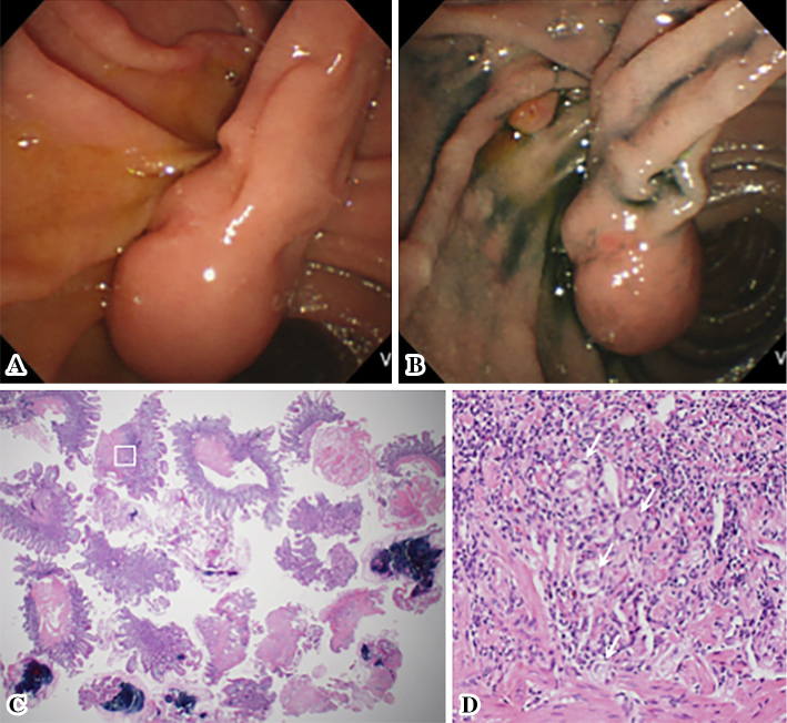
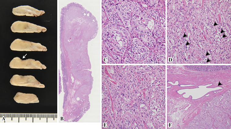
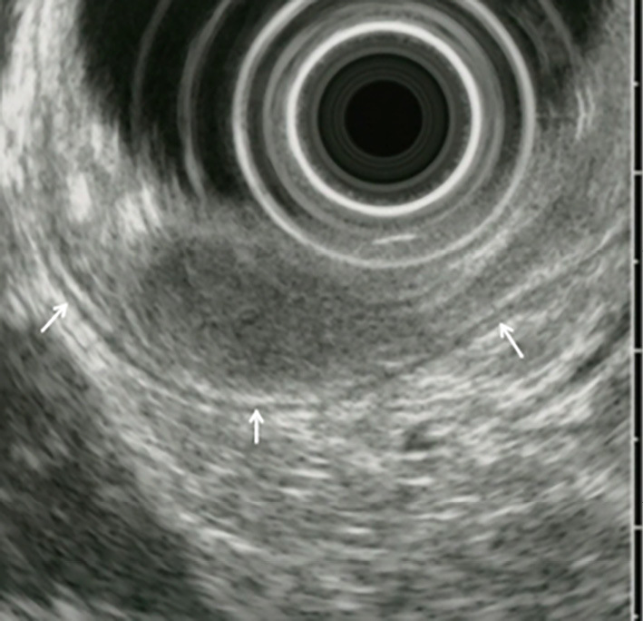
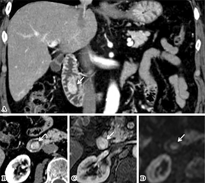
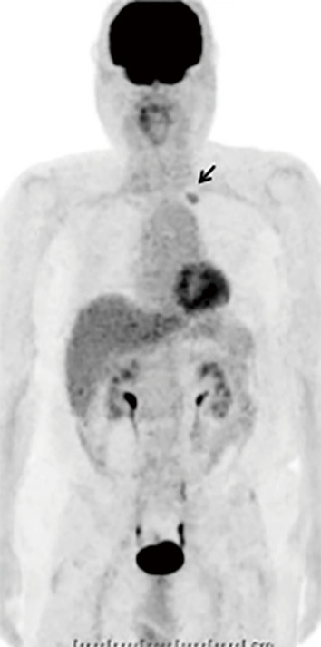
A duodenal polyp was found during a health check of a 71-year-old asymptomatic man. Duodenoscopy demonstrated a pedunculated, smooth-surfaced tumor of 18 mm in size, protruding from the minor papilla. Endoscopic ultrasonography demonstrated a homogeneously low-echoic submucosal tumor. Enhanced computed tomography and magnetic resonance imaging demonstrated a well-enhanced duodenal tumor without obvious metastasis. A tumor biopsy revealed a well-differentiated neuroendocrine tumor, and laparotomic transduodenal polypectomy with regional lymph node dissection was performed. The histology of the surgical specimen revealed gangliocytic paraganglioma consisting of three cell types: endocrine, ganglion, and spindle cells. There has been no recurrence in >5 years after surgery.
在一位71岁无症状男性的健康检查中发现了十二指肠息肉。十二指肠镜检查显示一个有蒂、表面光滑、大小为18毫米的肿瘤,从小乳头突出。内镜超声检查显示为均匀低回声的黏膜下肿瘤。增强计算机断层扫描和磁共振成像显示十二指肠肿瘤强化良好,无明显转移。肿瘤活检显示为高分化神经内分泌肿瘤,并进行了剖腹十二指肠息肉切除术及区域淋巴结清扫术。手术标本的组织学检查显示为神经节细胞性副神经节瘤,由三种细胞类型组成:内分泌细胞、神经节细胞和梭形细胞。术后5年多未复发。