• 文献检索
  • 文档翻译
  • 深度研究
  • 学术资讯
  • Suppr Zotero 插件Zotero 插件
  • 邀请有礼
  • 套餐&价格
  • 历史记录
应用&插件
Suppr Zotero 插件Zotero 插件浏览器插件Mac 客户端Windows 客户端微信小程序
定价
高级版会员购买积分包购买API积分包
服务
文献检索文档翻译深度研究API 文档MCP 服务
关于我们
关于 Suppr公司介绍联系我们用户协议隐私条款
关注我们

Suppr 超能文献

核心技术专利:CN118964589B侵权必究
粤ICP备2023148730 号-1Suppr @ 2026

文献检索

告别复杂PubMed语法,用中文像聊天一样搜索,搜遍4000万医学文献。AI智能推荐,让科研检索更轻松。

立即免费搜索

文件翻译

保留排版,准确专业,支持PDF/Word/PPT等文件格式,支持 12+语言互译。

免费翻译文档

深度研究

AI帮你快速写综述,25分钟生成高质量综述,智能提取关键信息,辅助科研写作。

立即免费体验

阿尔茨海默病患者的神经退行性变特征:一项脑形态测量学研究。

Neuro-degeneration profile of Alzheimer's patients: A brain morphometry study.

作者信息

Ramos Bernardes da Silva Filho Silvio, Oliveira Barbosa Jeam Haroldo, Rondinoni Carlo, Dos Santos Antonio Carlos, Garrido Salmon Carlos Ernesto, da Costa Lima Nereida Kilza, Ferriolli Eduardo, Moriguti Júlio César

机构信息

Ribeirao Preto Medical School of University of Sao Paulo, Ribeirao Preto, SP, Brazil.

Faculty of Philosophy, Sciences and Letters at Ribeirao Preto, University of Sao Paulo (FFCLRP-USP), Ribeirao Preto, SP, Brazil.

出版信息

Neuroimage Clin. 2017 Apr 3;15:15-24. doi: 10.1016/j.nicl.2017.04.001. eCollection 2017.

DOI:10.1016/j.nicl.2017.04.001
PMID:28459000
原文链接:https://pmc.ncbi.nlm.nih.gov/articles/PMC5397580/
Abstract

INTRODUCTION

Alzheimer's disease (AD) is a primary and progressive neurodegenerative disorder, which is marked by cognitive deterioration and memory impairment. Atrophy of hippocampus and other basal brain regions is one of the most predominant structural imaging findings related to AD. Most studies have evaluated the pre-clinical and initial stages of AD through clinical trials using Magnetic Resonance Imaging. Structural biomarkers for advanced AD stages have not been evaluated yet, being considered only hypothetically.

OBJECTIVE

To evaluate the brain morphometry of AD patients at all disease stages, identifying the structural neuro-degeneration profile associated with AD severity.

MATERIAL AND METHODS

AD patients aged 60 years or over at different AD stages were recruited and grouped into three groups following the Clinical Dementia Rating (CDR) score: CDR1 (n = 16), CDR2 (n = 15), CDR3 (n = 13). Age paired healthy volunteers (n = 16) were also recruited (control group). Brain images were acquired on a 3T magnetic resonance scanner using a conventional Gradient eco 3D T1-w sequence without contrast injection. Volumetric quantitative data and cortical thickness were obtained by automatic segmentation using the Freesurfer software. Volume of each brain region was normalized by the whole brain volume in order to minimize age and body size effects. Volume and cortical thickness variations among groups were compared.

RESULTS

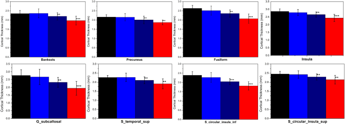
Atrophy was observed in the hippocampus, amygdala, entorhinal cortex, parahippocampal region, temporal pole and temporal lobe of patients suffering from AD at any stage. Cortical thickness was reduced only in the parahippocampal gyrus at all disease stages. Volume and cortical thickness were correlated with the Mini Mental State Examination (MMSE) score in all studied regions, as well as with CDR and disease duration.

DISCUSSION AND CONCLUSION

As previously reported, brain regions affected by AD during its initial stages, such as hippocampus, amygdala, entorhinal cortex, and parahippocampal region, were found to be altered even in individuals with severe AD. In addition, individuals, specifically, with CDR 3, have multiple regions with lower volumes than individuals with a CDR 2. These results indicate that rates of atrophy have not plateaued out at CDR 2-3, and in severe patients there are yet neuronal loss and gliosis. These findings can add important information to the more accepted model in the literature that focuses mainly on early stages. Our findings allow a better understanding on the AD pathophysiologic process and follow-up process of drug treatment even at advanced disease stages.

摘要

引言

阿尔茨海默病(AD)是一种原发性进行性神经退行性疾病,其特征为认知功能衰退和记忆障碍。海马体及其他脑基底区域萎缩是与AD相关的最主要结构影像学表现之一。大多数研究通过磁共振成像临床试验评估AD的临床前期和初始阶段。晚期AD阶段的结构生物标志物尚未得到评估,仅为假设性探讨。

目的

评估各疾病阶段AD患者的脑形态测量学特征,确定与AD严重程度相关的结构神经退行性变特征。

材料与方法

招募60岁及以上处于不同AD阶段的患者,根据临床痴呆评定量表(CDR)评分分为三组:CDR1组(n = 16)、CDR2组(n = 15)、CDR3组(n = 13)。还招募了年龄匹配的健康志愿者(n = 16)作为对照组。使用常规梯度回波3D T1加权序列在3T磁共振扫描仪上采集脑图像,不注射造影剂。通过Freesurfer软件自动分割获取体积定量数据和皮质厚度。每个脑区的体积通过全脑体积进行归一化,以尽量减少年龄和体型的影响。比较各组间的体积和皮质厚度变化。

结果

在任何阶段的AD患者中,均观察到海马体、杏仁核、内嗅皮质、海马旁区域、颞极和颞叶萎缩。在所有疾病阶段,仅海马旁回皮质厚度降低。在所有研究区域,体积和皮质厚度均与简易精神状态检查表(MMSE)评分相关,也与CDR和病程相关。

讨论与结论

如先前报道,AD早期受影响的脑区,如海马体、杏仁核、内嗅皮质和海马旁区域,即使在重度AD个体中也有改变。此外,特别是CDR 3的个体,与CDR 2的个体相比,有多个脑区体积更小。这些结果表明,萎缩率在CDR 2 - 3阶段并未趋于平稳,在重度患者中仍存在神经元丢失和胶质增生。这些发现可为文献中主要关注早期阶段的更普遍接受的模型增添重要信息。我们的发现有助于更好地理解AD的病理生理过程以及即使在疾病晚期的药物治疗随访过程。

https://cdn.ncbi.nlm.nih.gov/pmc/blobs/4291/5397580/3fe387f4ff9d/gr10.jpg
https://cdn.ncbi.nlm.nih.gov/pmc/blobs/4291/5397580/19581db318b8/gr1.jpg
https://cdn.ncbi.nlm.nih.gov/pmc/blobs/4291/5397580/a8659581a314/gr2.jpg
https://cdn.ncbi.nlm.nih.gov/pmc/blobs/4291/5397580/65f4bc1e7498/gr3.jpg
https://cdn.ncbi.nlm.nih.gov/pmc/blobs/4291/5397580/ba7419cdafe3/gr4.jpg
https://cdn.ncbi.nlm.nih.gov/pmc/blobs/4291/5397580/00791b7605bb/gr5.jpg
https://cdn.ncbi.nlm.nih.gov/pmc/blobs/4291/5397580/82547c0912f9/gr6.jpg
https://cdn.ncbi.nlm.nih.gov/pmc/blobs/4291/5397580/0dede6068fdb/gr7.jpg
https://cdn.ncbi.nlm.nih.gov/pmc/blobs/4291/5397580/b5c24a43e18b/gr8.jpg
https://cdn.ncbi.nlm.nih.gov/pmc/blobs/4291/5397580/9609e84f1c38/gr9.jpg
https://cdn.ncbi.nlm.nih.gov/pmc/blobs/4291/5397580/3fe387f4ff9d/gr10.jpg

相似文献

1
Neuro-degeneration profile of Alzheimer's patients: A brain morphometry study.阿尔茨海默病患者的神经退行性变特征:一项脑形态测量学研究。
Neuroimage Clin. 2017 Apr 3;15:15-24. doi: 10.1016/j.nicl.2017.04.001. eCollection 2017.
2
Structural magnetic resonance imaging for the early diagnosis of dementia due to Alzheimer's disease in people with mild cognitive impairment.用于轻度认知障碍患者阿尔茨海默病所致痴呆早期诊断的结构磁共振成像
Cochrane Database Syst Rev. 2020 Mar 2;3(3):CD009628. doi: 10.1002/14651858.CD009628.pub2.
3
Longitudinal structural cerebral changes related to core CSF biomarkers in preclinical Alzheimer's disease: A study of two independent datasets.与临床前阿尔茨海默病核心 CSF 生物标志物相关的纵向结构脑变化:两项独立数据集研究。
Neuroimage Clin. 2018 Apr 16;19:190-201. doi: 10.1016/j.nicl.2018.04.016. eCollection 2018.
4
The influence of hippocampal atrophy on the cognitive phenotype of dementia with Lewy bodies.海马体萎缩对路易体痴呆认知表型的影响。
Int J Geriatr Psychiatry. 2017 Nov;32(11):1182-1189. doi: 10.1002/gps.4719. Epub 2017 Apr 20.
5
Structural Alteration of Medial Temporal Lobe Subfield in the Amnestic Mild Cognitive Impairment Stage of Alzheimer's Disease.内侧颞叶亚区结构改变在阿尔茨海默病遗忘型轻度认知障碍阶段。
Neural Plast. 2022 Jan 24;2022:8461235. doi: 10.1155/2022/8461235. eCollection 2022.
6
Associations of tau, Aβ, and brain volume of the Papez circuit with cognition in Alzheimer's disease.阿尔茨海默病中tau蛋白、β淀粉样蛋白以及帕佩兹环路脑容量与认知的关联。
Eur Arch Psychiatry Clin Neurosci. 2025 Feb;275(1):241-256. doi: 10.1007/s00406-024-01827-7. Epub 2024 Jun 2.
7
In vivo detection of microstructural correlates of brain pathology in preclinical and early Alzheimer Disease with magnetic resonance imaging.利用磁共振成像在临床前和早期阿尔茨海默病中对脑病理微观结构关联进行体内检测。
Neuroimage. 2017 Mar 1;148:296-304. doi: 10.1016/j.neuroimage.2016.12.026. Epub 2016 Dec 15.
8
Patterns of Grey Matter Atrophy at Different Stages of Parkinson's and Alzheimer's Diseases and Relation to Cognition.帕金森病和阿尔茨海默病不同阶段的灰质萎缩模式及其与认知的关系。
Brain Topogr. 2019 Jan;32(1):142-160. doi: 10.1007/s10548-018-0675-2. Epub 2018 Sep 11.
9
Quantitative Gradient Echo MRI Identifies Dark Matter as a New Imaging Biomarker of Neurodegeneration that Precedes Tisssue Atrophy in Early Alzheimer's Disease.定量梯度回波 MRI 将暗物质鉴定为神经退行性变的新成像生物标志物,可在早期阿尔茨海默病中出现组织萎缩之前预测疾病。
J Alzheimers Dis. 2022;85(2):905-924. doi: 10.3233/JAD-210503.
10
Cross-Sectional Comparison of Structural MRI Markers of Impairment in a Diverse Cohort of Older Adults.不同老年人群体中损伤的结构磁共振成像标志物的横断面比较
Hum Brain Mapp. 2025 Feb 1;46(2):e70133. doi: 10.1002/hbm.70133.

引用本文的文献

1
The Effect of Overcoming the Digital Divide on Middle Frontal Gyrus Atrophy in Aging Adults: Large-Scale Retrospective Magnetic Resonance Imaging Cohort Study.克服数字鸿沟对老年人额中回萎缩的影响:大规模回顾性磁共振成像队列研究。
J Med Internet Res. 2025 Jul 22;27:e73360. doi: 10.2196/73360.
2
Bioinformatic Analysis of Alzheimer's Disease Metabolomic Datasets for Ceramides and Gangliosides.用于神经酰胺和神经节苷脂的阿尔茨海默病代谢组学数据集的生物信息学分析
Methods Mol Biol. 2025;2925:407-412. doi: 10.1007/978-1-0716-4534-5_27.
3
Increasing H2B Monoubiquitination Improves the Transcriptome and Memory in the Aged Hippocampus.

本文引用的文献

1
Diagnosis of Alzheimer's disease in Brazil: Supplementary exams.巴西阿尔茨海默病的诊断:补充检查
Dement Neuropsychol. 2011 Jul-Sep;5(3):167-177. doi: 10.1590/S1980-57642011DN05030004.
2
Cortical Thickness in Dementia with Lewy Bodies and Alzheimer's Disease: A Comparison of Prodromal and Dementia Stages.路易体痴呆和阿尔茨海默病的皮质厚度:前驱期与痴呆期的比较
PLoS One. 2015 Jun 10;10(6):e0127396. doi: 10.1371/journal.pone.0127396. eCollection 2015.
3
Advancing research diagnostic criteria for Alzheimer's disease: the IWG-2 criteria.
增加H2B单泛素化可改善老年海马体的转录组和记忆力。
eNeuro. 2025 Apr 11;12(4). doi: 10.1523/ENEURO.0037-25.2025. Print 2025 Apr.
4
Exploring the Potential of tsRNA as Biomarkers for Diagnosis and Treatment of Neurogenetic Disorders.探索转运RNA衍生的小RNA作为神经遗传性疾病诊断和治疗生物标志物的潜力。
Mol Neurobiol. 2025 Feb 26. doi: 10.1007/s12035-025-04760-5.
5
Exploring the versatility of as a model organism in biomedical research: a comprehensive review.探索[未提及具体生物]作为生物医学研究模式生物的多功能性:一项综合综述。 (注:原文中“Exploring the versatility of as a model organism”这里缺少具体所指生物)
Fly (Austin). 2025 Dec;19(1):2420453. doi: 10.1080/19336934.2024.2420453. Epub 2024 Dec 25.
6
Iron accumulation/overload and Alzheimer's disease risk factors in the precuneus region: A comprehensive narrative review.楔前叶区域的铁蓄积/过载与阿尔茨海默病风险因素:一项全面的叙述性综述。
Aging Med (Milton). 2024 Oct 22;7(5):649-667. doi: 10.1002/agm2.12363. eCollection 2024 Oct.
7
Morphological characterization of the hippocampus: a first database in Ecuador.海马体的形态学特征:厄瓜多尔的首个数据库。
Front Hum Neurosci. 2024 Oct 18;18:1387212. doi: 10.3389/fnhum.2024.1387212. eCollection 2024.
8
Exploring the Role of Reactive Oxygen Species in the Pathogenesis and Pathophysiology of Alzheimer's and Parkinson's Disease and the Efficacy of Antioxidant Treatment.探索活性氧在阿尔茨海默病和帕金森病发病机制及病理生理学中的作用以及抗氧化治疗的疗效。
Antioxidants (Basel). 2024 Sep 20;13(9):1138. doi: 10.3390/antiox13091138.
9
The association between rs6859 in gene and Alzheimer's disease is partly mediated by pTau.基因中rs6859与阿尔茨海默病之间的关联部分由磷酸化tau蛋白介导。
Front Aging Neurosci. 2024 Aug 6;16:1388363. doi: 10.3389/fnagi.2024.1388363. eCollection 2024.
10
The association between rs6859 in gene and Alzheimer's disease is partly mediated by pTau.基因中rs6859与阿尔茨海默病之间的关联部分是由磷酸化tau蛋白介导的。
medRxiv. 2024 Jun 22:2024.06.21.24309310. doi: 10.1101/2024.06.21.24309310.
推进阿尔茨海默病研究诊断标准:IWG-2 标准。
Lancet Neurol. 2014 Jun;13(6):614-29. doi: 10.1016/S1474-4422(14)70090-0.
4
Assessment of regional MR diffusion changes in dementia with Lewy bodies and Alzheimer's disease.路易体痴呆和阿尔茨海默病中区域磁共振扩散变化的评估。
Int Psychogeriatr. 2014 Apr;26(4):627-35. doi: 10.1017/S1041610213002317. Epub 2013 Dec 16.
5
What causes alzheimer's disease?阿尔茨海默病的病因是什么?
Folia Neuropathol. 2013;51(3):169-88. doi: 10.5114/fn.2013.37702.
6
Sensitivity of different MRI-techniques to assess gray matter atrophy patterns in Alzheimer's disease is region-specific.不同 MRI 技术评估阿尔茨海默病灰质萎缩模式的敏感性具有区域特异性。
Curr Alzheimer Res. 2013 Nov;10(9):940-51. doi: 10.2174/15672050113109990158.
7
Tracking pathophysiological processes in Alzheimer's disease: an updated hypothetical model of dynamic biomarkers.阿尔茨海默病病理生理过程的追踪:动态生物标志物的更新假设模型。
Lancet Neurol. 2013 Feb;12(2):207-16. doi: 10.1016/S1474-4422(12)70291-0.
8
An MRI-based index to measure the severity of Alzheimer's disease-like structural pattern in subjects with mild cognitive impairment.基于 MRI 的指标,用于衡量轻度认知障碍患者阿尔茨海默病样结构模式的严重程度。
J Intern Med. 2013 Apr;273(4):396-409. doi: 10.1111/joim.12028. Epub 2013 Jan 30.
9
Cognitive reserve in ageing and Alzheimer's disease.衰老和阿尔茨海默病中的认知储备。
Lancet Neurol. 2012 Nov;11(11):1006-12. doi: 10.1016/S1474-4422(12)70191-6.
10
Shapes of the trajectories of 5 major biomarkers of Alzheimer disease.阿尔茨海默病5种主要生物标志物的轨迹形状。
Arch Neurol. 2012 Jul;69(7):856-67. doi: 10.1001/archneurol.2011.3405.