Jin Chengbiao, Lee Gayoung, Oh Changseok, Kim Hyun Jung, Kim Hyun-Man
Laboratory for the Study of Molecular Biointerfaces, Department of Oral Histology and Developmental Biology, Program of Cell and Developmental Biology, Dental Research Institute, Seoul National University School of Dentistry, Seoul, Korea.
J Periodontal Implant Sci. 2017 Apr;47(2):116-131. doi: 10.5051/jpis.2017.47.2.116. Epub 2017 Apr 29.
The entry of bacteria or harmful substances through the epithelial seal of human gingival keratinocytes (HGKs) in the junctional epithelium (JE) is blocked by specialized intercellular junctions such as E-cadherin junctions (ECJs). However, the influence of roughened substrates, which may occur due to apical migration of the JE, root planing, or peri-implantitis, on the development of the ECJs of HGKs remains largely unknown.
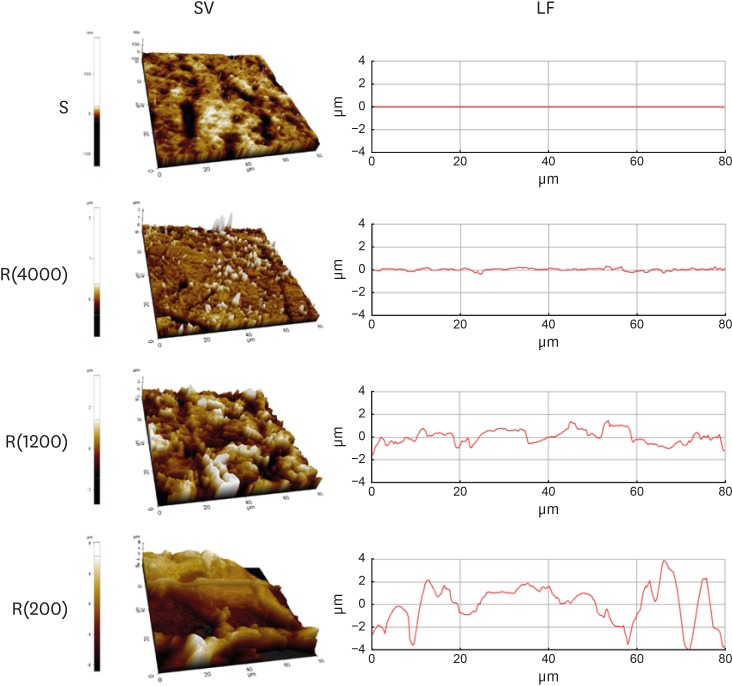
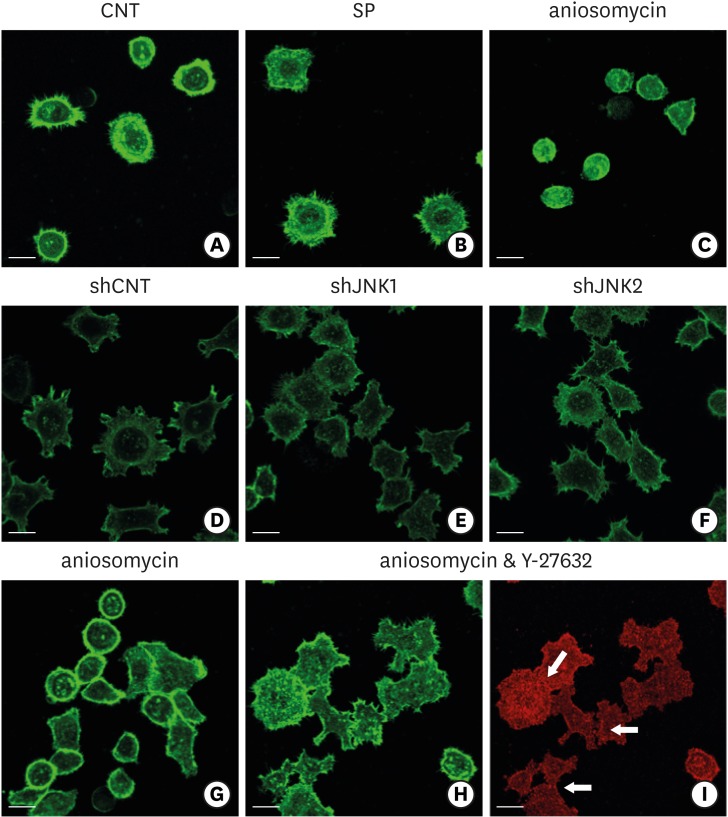
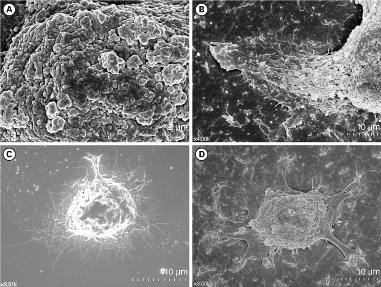
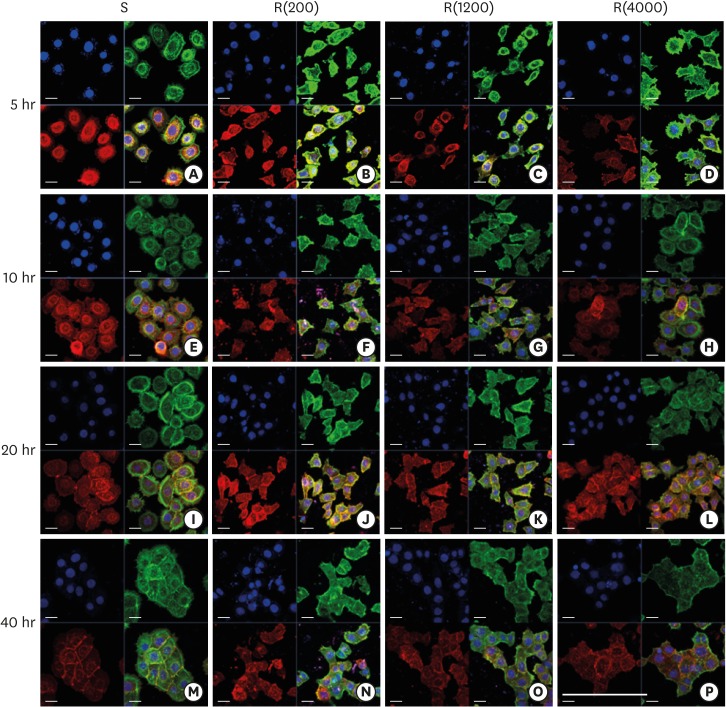
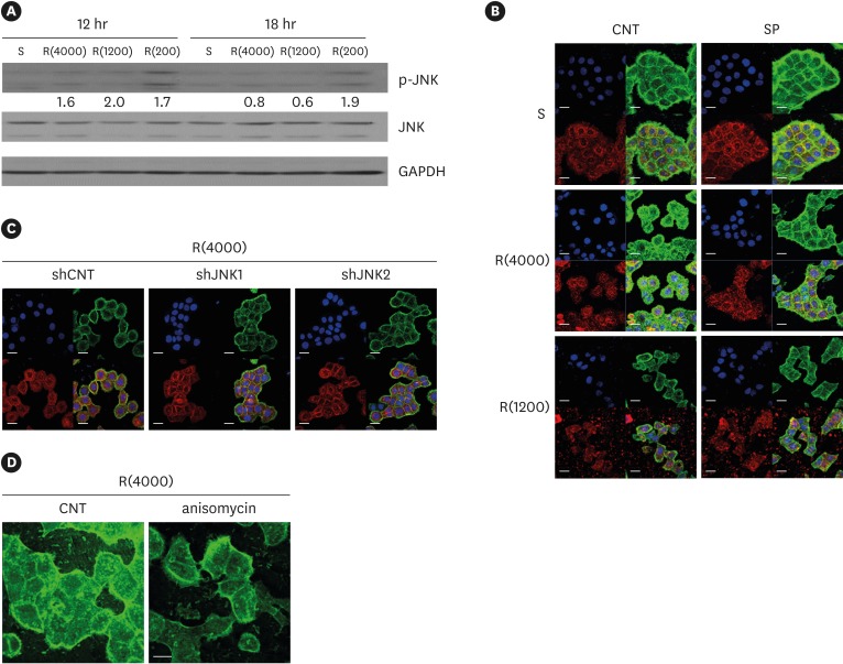
HGKs were cultured on substrates with varying levels of roughness, which were prepared by rubbing hydrophobic polystyrene dishes with silicon carbide papers. The activity of c-Jun N-terminal kinase (JNK) was inhibited with SP600125 or by transfection with JNK short hairpin RNA. The development of intercellular junctions was analyzed using scanning electron microscopy or confocal laser scanning microscopy after immunohistochemical staining of the cells for E-cadherin. The expression level of phospho-JNK was assessed by immunoblotting.
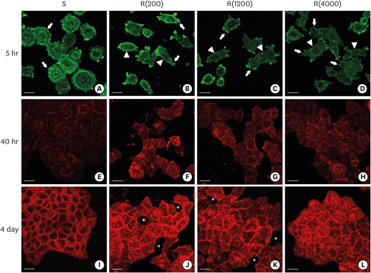
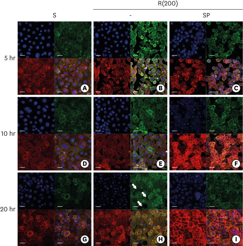
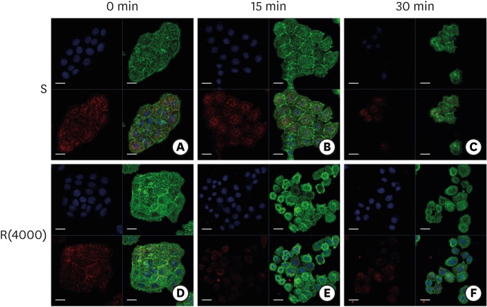
HGKs developed tight intercellular junctions devoid of wide intercellular gaps on smooth substrates and on rough substrates with low-nanometer dimensions (average roughness []=121.3±13.4 nm), although the ECJs of HGKs on rough substrates with low-nanometer dimensions developed later than those of HGKs on smooth substrates. In contrast, HGKs developed short intercellular junctions with wide intercellular gaps on rough substrates with mid- or high-nanometer dimensions (=505.3±115.3 nm, 867.0±168.6 nm). Notably, the stability of the ECJs was low on the rough substrates, as demonstrated by the rapid destruction of the cell junction following calcium depletion. Inhibition of JNK activity promoted ECJ development in HGKs. JNK was closely associated with cortical actin in the regulation of ECJs in HGKs.
These results indicate that on rough substrates with nanometer dimensions, the ECJs of HGKs develop slowly or defectively, and that this effect can be reversed by inhibiting JNK.
细菌或有害物质通过结合上皮(JE)中人类牙龈角质形成细胞(HGK)的上皮封闭进入体内,会被诸如E-钙黏蛋白连接(ECJ)等特殊的细胞间连接所阻断。然而,由于JE的根尖迁移、根面平整或种植体周围炎可能导致的粗糙底物,对HGK的ECJ发育的影响在很大程度上仍不清楚。
将HGK培养在具有不同粗糙度水平的底物上,这些底物是通过用碳化硅纸摩擦疏水聚苯乙烯培养皿制备的。用SP600125或通过转染JNK短发夹RNA抑制c-Jun氨基末端激酶(JNK)的活性。在对细胞进行E-钙黏蛋白免疫组织化学染色后,使用扫描电子显微镜或共聚焦激光扫描显微镜分析细胞间连接的发育情况。通过免疫印迹评估磷酸化JNK的表达水平。
HGK在光滑底物和具有低纳米尺寸(平均粗糙度[]=121.3±13.4 nm)的粗糙底物上形成紧密的细胞间连接,没有宽的细胞间隙,尽管HGK在具有低纳米尺寸的粗糙底物上的ECJ比在光滑底物上的HGK发育得晚。相比之下,HGK在具有中或高纳米尺寸(=505.3±115.3 nm,867.0±168.6 nm)的粗糙底物上形成具有宽细胞间隙的短细胞间连接。值得注意的是,在粗糙底物上ECJ的稳定性较低,这通过钙耗尽后细胞连接的快速破坏得以证明。抑制JNK活性促进了HGK中ECJ的发育。在HGK的ECJ调节中,JNK与皮质肌动蛋白密切相关。
这些结果表明,在具有纳米尺寸的粗糙底物上,HGK的ECJ发育缓慢或存在缺陷,并且这种影响可以通过抑制JNK来逆转。