• 文献检索
  • 文档翻译
  • 深度研究
  • 学术资讯
  • Suppr Zotero 插件Zotero 插件
  • 邀请有礼
  • 套餐&价格
  • 历史记录
应用&插件
Suppr Zotero 插件Zotero 插件浏览器插件Mac 客户端Windows 客户端微信小程序
定价
高级版会员购买积分包购买API积分包
服务
文献检索文档翻译深度研究API 文档MCP 服务
关于我们
关于 Suppr公司介绍联系我们用户协议隐私条款
关注我们

Suppr 超能文献

核心技术专利:CN118964589B侵权必究
粤ICP备2023148730 号-1Suppr @ 2026

文献检索

告别复杂PubMed语法,用中文像聊天一样搜索,搜遍4000万医学文献。AI智能推荐,让科研检索更轻松。

立即免费搜索

文件翻译

保留排版,准确专业,支持PDF/Word/PPT等文件格式,支持 12+语言互译。

免费翻译文档

深度研究

AI帮你快速写综述,25分钟生成高质量综述,智能提取关键信息,辅助科研写作。

立即免费体验

JNK 信号转导作为软连接组织生理学、病理学和愈合的关键调节剂。

JNK Signaling as a Key Modulator of Soft Connective Tissue Physiology, Pathology, and Healing.

机构信息

Department of Anatomy & Cell Biology, Schulich School of Medicine and Dentistry, University of Western Ontario, 1151 Richmond St, London, ON N6A 5C1, Canada.

Biomedical Engineering Graduate Program, Schulich School of Medicine and Dentistry, University of Western Ontario, 1151 Richmond St, London, ON N6A 5C1, Canada.

出版信息

Int J Mol Sci. 2020 Feb 4;21(3):1015. doi: 10.3390/ijms21031015.

DOI:10.3390/ijms21031015
PMID:32033060
原文链接:https://pmc.ncbi.nlm.nih.gov/articles/PMC7037145/
Abstract

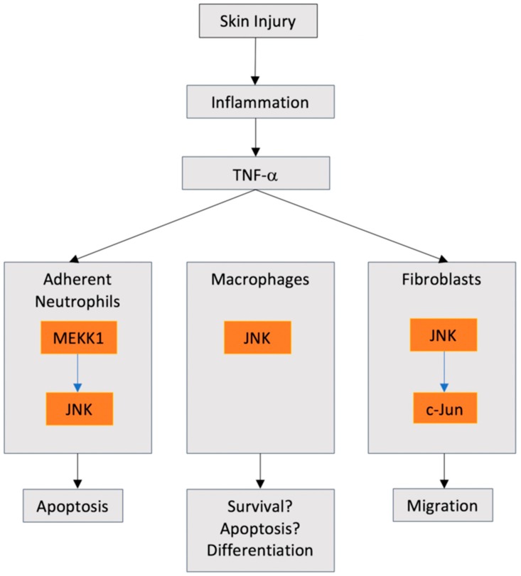
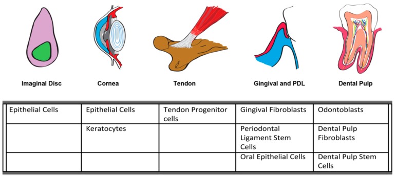
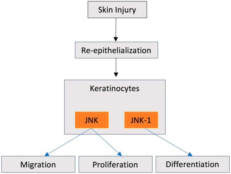
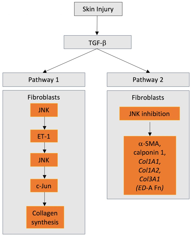
In healthy individuals, the healing of soft tissues such as skin after pathological insult or post injury follows a relatively predictable and defined series of cell and molecular processes to restore tissue architecture and function(s). Healing progresses through the phases of hemostasis, inflammation, proliferation, remodeling, and concomitant with re-epithelialization restores barrier function. Soft tissue healing is achieved through the spatiotemporal interplay of multiple different cell types including neutrophils, monocytes/macrophages, fibroblasts, endothelial cells/pericytes, and keratinocytes. Expressed in most cell types, c-Jun N-terminal kinases (JNK) are signaling molecules associated with the regulation of several cellular processes involved in soft tissue wound healing and in response to cellular stress. A member of the mitogen-activated protein kinase family (MAPK), JNKs have been implicated in the regulation of inflammatory cell phenotype, as well as fibroblast, stem/progenitor cell, and epithelial cell biology. In this review, we discuss our understanding of JNKs in the regulation of cell behaviors related to tissue injury, pathology, and wound healing of soft tissues. Using models as diverse as , mice, rats, as well as human tissues, research is now defining important, but sometimes conflicting roles for JNKs in the regulation of multiple molecular processes in multiple different cell types central to wound healing processes. In this review, we focus specifically on the role of JNKs in the regulation of cell behavior in the healing of skin, cornea, tendon, gingiva, and dental pulp tissues. We conclude that while parallels can be drawn between some JNK activities and the control of cell behavior in healing, the roles of JNK can also be very specific modes of action depending on the tissue and the phase of healing.

摘要

在健康个体中,皮肤等软组织在病理性损伤或创伤后愈合遵循相对可预测和明确的细胞和分子过程系列,以恢复组织结构和功能。愈合通过止血、炎症、增殖、重塑等阶段进行,同时伴随着再上皮化恢复屏障功能。软组织愈合是通过多种不同细胞类型(包括中性粒细胞、单核细胞/巨噬细胞、成纤维细胞、内皮细胞/周细胞和角质形成细胞)的时空相互作用实现的。c-Jun N 末端激酶(JNK)在大多数细胞类型中表达,是与调节参与软组织伤口愈合的几种细胞过程以及对细胞应激的反应相关的信号分子。JNK 是丝裂原激活蛋白激酶家族(MAPK)的成员,与调节炎症细胞表型以及成纤维细胞、干细胞/祖细胞和上皮细胞生物学有关。在这篇综述中,我们讨论了我们对 JNK 在调节与组织损伤、病理学和软组织伤口愈合相关的细胞行为的理解。使用范围广泛的模型,如小鼠、大鼠以及人类组织,研究现在正在定义 JNK 在调节与伤口愈合过程中多种不同细胞类型的多种分子过程相关的细胞行为方面的重要但有时相互矛盾的作用。在这篇综述中,我们特别关注 JNK 在调节皮肤、角膜、肌腱、牙龈和牙髓组织愈合过程中细胞行为的作用。我们的结论是,虽然可以在某些 JNK 活性和对愈合中细胞行为的控制之间得出一些相似之处,但 JNK 的作用也可以根据组织和愈合阶段而具有非常具体的作用模式。

https://cdn.ncbi.nlm.nih.gov/pmc/blobs/926e/7037145/45d31308bac6/ijms-21-01015-g005.jpg
https://cdn.ncbi.nlm.nih.gov/pmc/blobs/926e/7037145/8a4ce8d5bdc7/ijms-21-01015-g001.jpg
https://cdn.ncbi.nlm.nih.gov/pmc/blobs/926e/7037145/8930ce0b09ef/ijms-21-01015-g002.jpg
https://cdn.ncbi.nlm.nih.gov/pmc/blobs/926e/7037145/bea199d424aa/ijms-21-01015-g003.jpg
https://cdn.ncbi.nlm.nih.gov/pmc/blobs/926e/7037145/0fdac972c125/ijms-21-01015-g004.jpg
https://cdn.ncbi.nlm.nih.gov/pmc/blobs/926e/7037145/45d31308bac6/ijms-21-01015-g005.jpg

相似文献

1
JNK Signaling as a Key Modulator of Soft Connective Tissue Physiology, Pathology, and Healing.JNK 信号转导作为软连接组织生理学、病理学和愈合的关键调节剂。
Int J Mol Sci. 2020 Feb 4;21(3):1015. doi: 10.3390/ijms21031015.
2
The activation of the NF-κB-JNK pathway is independent of the PI3K-Rac1-JNK pathway involved in the bFGF-regulated human fibroblast cell migration.NF-κB-JNK信号通路的激活独立于参与碱性成纤维细胞生长因子(bFGF)调节的人成纤维细胞迁移的PI3K-Rac1-JNK信号通路。
J Dermatol Sci. 2016 Apr;82(1):28-37. doi: 10.1016/j.jdermsci.2016.01.003. Epub 2016 Jan 7.
3
JNK MAPK signaling contributes in vivo to injury-induced corneal epithelial migration.JNK MAPK 信号通路在体内有助于损伤诱导的角膜上皮迁移。
Ophthalmic Res. 2009;42(4):185-92. doi: 10.1159/000232401. Epub 2009 Aug 7.
4
Galloyl benzamide-based compounds modulating tumour necrosis factor α-stimulated c-Jun N-terminal kinase and p38 mitogen-activated protein kinase signalling pathways.基于没食子酰苯甲酰胺的化合物调节肿瘤坏死因子α刺激的c-Jun N端激酶和p38丝裂原活化蛋白激酶信号通路。
J Pharm Pharmacol. 2015 Oct;67(10):1380-92. doi: 10.1111/jphp.12438. Epub 2015 Jun 16.
5
Expression of the Alternative Oxidase Influences Jun N-Terminal Kinase Signaling and Cell Migration.交替氧化酶的表达影响 Jun N-末端激酶信号转导和细胞迁移。
Mol Cell Biol. 2018 Nov 28;38(24). doi: 10.1128/MCB.00110-18. Print 2018 Dec 15.
6
Role of Jun N-terminal Kinase (JNK) signaling in the wound healing and regeneration of a Drosophila melanogaster wing imaginal disc.JNK信号通路在黑腹果蝇翅成虫盘伤口愈合和再生中的作用
Int J Dev Biol. 2005;49(4):391-9. doi: 10.1387/ijdb.052006jm.
7
CD9 is critical for cutaneous wound healing through JNK signaling.CD9 通过 JNK 信号通路对皮肤伤口愈合起关键作用。
J Invest Dermatol. 2012 Jan;132(1):226-36. doi: 10.1038/jid.2011.268. Epub 2011 Sep 1.
8
JNK signaling pathway is required for efficient wound healing in Drosophila.JNK信号通路在果蝇高效伤口愈合过程中是必需的。
Dev Biol. 2002 Jan 1;241(1):145-56. doi: 10.1006/dbio.2001.0502.
9
Efficient keratinocyte differentiation strictly depends on JNK-induced soluble factors in fibroblasts.成纤维细胞中 JNK 诱导的可溶性因子严格依赖于角质形成细胞的有效分化。
J Invest Dermatol. 2014 May;134(5):1332-1341. doi: 10.1038/jid.2013.535. Epub 2013 Nov 16.
10
Activation of JNK signaling mediates connective tissue growth factor expression and scar formation in corneal wound healing.JNK 信号的激活介导了角膜伤口愈合过程中结缔组织生长因子的表达和瘢痕形成。
PLoS One. 2012;7(2):e32128. doi: 10.1371/journal.pone.0032128. Epub 2012 Feb 21.

引用本文的文献

1
Biomolecule-functionalized dental implant surfaces: Towards augmenting soft tissue integration.生物分子功能化的牙种植体表面:促进软组织整合
Bioact Mater. 2025 Jul 26;53:540-590. doi: 10.1016/j.bioactmat.2025.07.005. eCollection 2025 Nov.
2
Colostrum-Derived Exosomal Lactoferrin Promotes Skin Fibroblast Regeneration by Suppressing Inflammatory Responses.初乳来源的外泌体乳铁蛋白通过抑制炎症反应促进皮肤成纤维细胞再生。
Curr Issues Mol Biol. 2025 Jul 15;47(7):549. doi: 10.3390/cimb47070549.
3
Role of C-Jun N-Terminal Kinases on a Stressed Epithelium: Time for Testing Isoform Specificity.

本文引用的文献

1
Non-canonical (non-SMAD2/3) TGF-β signaling in fibrosis: Mechanisms and targets.非经典(非 SMAD2/3)TGF-β 信号通路在纤维化中的作用:机制与靶点。
Semin Cell Dev Biol. 2020 May;101:115-122. doi: 10.1016/j.semcdb.2019.11.013. Epub 2019 Dec 26.
2
Aspirin inhibits inflammation and scar formation in the injury tendon healing through regulating JNK/STAT-3 signalling pathway.阿司匹林通过调节 JNK/STAT-3 信号通路抑制损伤肌腱愈合中的炎症和瘢痕形成。
Cell Prolif. 2019 Jul;52(4):e12650. doi: 10.1111/cpr.12650. Epub 2019 Jun 21.
3
Loss of Wnt4 expression inhibits the odontogenic potential of dental pulp stem cells through JNK signaling in pulpitis.
C-Jun氨基末端激酶在应激上皮中的作用:检验亚型特异性的时候到了。
Biology (Basel). 2025 Jun 3;14(6):649. doi: 10.3390/biology14060649.
4
GMP manufacturing of umbilical cord blood mesenchymal stem cell-released exosomes and verification of wound healing efficacy.脐带血间充质干细胞释放的外泌体的GMP生产及伤口愈合功效验证。
Nanotheranostics. 2025 Mar 3;9(2):95-109. doi: 10.7150/ntno.105433. eCollection 2025.
5
Single-Cell Multiomics Analysis of Early Wound Response Programs in the Mouse Corneal Epithelium.小鼠角膜上皮早期伤口反应程序的单细胞多组学分析
Invest Ophthalmol Vis Sci. 2025 Mar 3;66(3):9. doi: 10.1167/iovs.66.3.9.
6
Revisited and innovative perspectives of oral ulcer: from biological specificity to local treatment.口腔溃疡的再审视与创新观点:从生物学特异性到局部治疗
Front Bioeng Biotechnol. 2024 Feb 22;12:1335377. doi: 10.3389/fbioe.2024.1335377. eCollection 2024.
7
Differential Effects of Overexpression of Wild Type and Kinase-Dead MELK in Fibroblasts and Keratinocytes, Potential Implications for Skin Wound Healing and Cancer.野生型和激酶失活型 MELK 在成纤维细胞和角质细胞中的差异表达及其对皮肤创伤愈合和癌症的潜在影响。
Int J Mol Sci. 2023 Apr 30;24(9):8089. doi: 10.3390/ijms24098089.
8
A Novel (L.) Britton Cell-Derived Phytocomplex Regulates Keratinocytes Inflammatory Cascade and Barrier Function and Preserves Vaginal Mucosal Integrity In Vivo.一种新型的源于(L.)布里顿细胞的植物复合物在体内调节角质形成细胞的炎症级联反应和屏障功能,并维持阴道黏膜完整性。
Pharmaceutics. 2023 Jan 10;15(1):240. doi: 10.3390/pharmaceutics15010240.
9
Pathology of Initial Axon Segments in Chronic Inflammatory Demyelinating Polyradiculoneuropathy and Related Disorders.慢性炎症性脱髓鞘性多发性神经病及相关疾病中初始轴突节段的病理学。
Int J Mol Sci. 2022 Nov 7;23(21):13621. doi: 10.3390/ijms232113621.
10
Fibroblasts in Scar Formation: Biology and Clinical Translation.瘢痕形成中的成纤维细胞:生物学与临床转化。
Oxid Med Cell Longev. 2022 May 12;2022:4586569. doi: 10.1155/2022/4586569. eCollection 2022.
Wnt4表达缺失通过牙髓炎症中的JNK信号传导抑制牙髓干细胞的成牙潜能。
Am J Transl Res. 2019 Mar 15;11(3):1819-1826. eCollection 2019.
4
: A Model Organism to Study Cancer.一种用于研究癌症的模式生物。
Front Genet. 2019 Mar 1;10:51. doi: 10.3389/fgene.2019.00051. eCollection 2019.
5
Fibronectin synthesis, but not α-smooth muscle expression, is regulated by periostin in gingival healing through FAK/JNK signaling.在牙龈愈合过程中,骨膜蛋白通过 FAK/JNK 信号通路调节纤连蛋白的合成,但不调节α-平滑肌肌动蛋白的表达。
Sci Rep. 2019 Feb 25;9(1):2708. doi: 10.1038/s41598-018-35805-6.
6
High Glucose Suppresses Keratinocyte Migration Through the Inhibition of p38 MAPK/Autophagy Pathway.高糖通过抑制p38丝裂原活化蛋白激酶/自噬途径抑制角质形成细胞迁移。
Front Physiol. 2019 Jan 28;10:24. doi: 10.3389/fphys.2019.00024. eCollection 2019.
7
Periostin and CCN2 Scaffolds Promote the Wound Healing Response in the Skin of Diabetic Mice.骨膜蛋白和 CCN2 支架促进糖尿病小鼠皮肤的伤口愈合反应。
Tissue Eng Part A. 2019 Sep;25(17-18):1326-1339. doi: 10.1089/ten.TEA.2018.0268. Epub 2019 Feb 27.
8
Resveratrol represses tumor necrosis factor α/c-Jun N-terminal kinase signaling via autophagy in human dental pulp stem cells.白藜芦醇通过自噬抑制人牙髓干细胞中肿瘤坏死因子 α/c-Jun N-末端激酶信号通路。
Arch Oral Biol. 2019 Jan;97:116-121. doi: 10.1016/j.archoralbio.2018.10.020. Epub 2018 Oct 22.
9
Crosstalk between mitogen-activated protein kinase inhibitors and transforming growth factor-β signaling results in variable activation of human dermal fibroblasts.丝裂原活化蛋白激酶抑制剂与转化生长因子-β信号通路的相互作用导致人真皮成纤维细胞的激活情况出现差异。
Int J Mol Med. 2019 Jan;43(1):325-335. doi: 10.3892/ijmm.2018.3949. Epub 2018 Oct 23.
10
Mechanistic role of transglutaminase-2 in focal adhesions.转谷氨酰胺酶-2 在黏着斑中的机械作用。
Sci Rep. 2018 Aug 17;8(1):12370. doi: 10.1038/s41598-018-30172-8.