Li Jingjing, Miao Lianjie, Zhao Chen, Shaikh Qureshi Wasay Mohiuddin, Shieh David, Guo Hua, Lu Yangyang, Hu Saiyang, Huang Alice, Zhang Lu, Cai Chen-Leng, Wan Leo Q, Xin Hongbo, Vincent Peter, Singer Harold A, Zheng Yi, Cleaver Ondine, Fan Zhen-Chuan, Wu Mingfu
Department of Molecular and Cellular Physiology, Albany Medical College, Albany, NY 12208, USA.
Institute of Translational Medicine, Nanchang University, Nanchang 330031, China.
Development. 2017 May 1;144(9):1635-1647. doi: 10.1242/dev.147173.
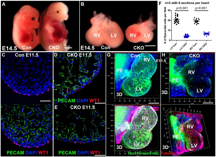
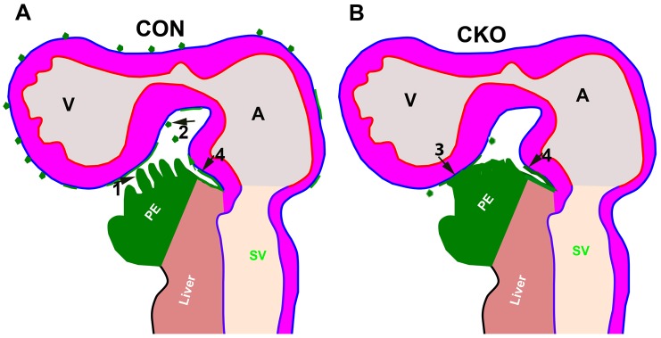
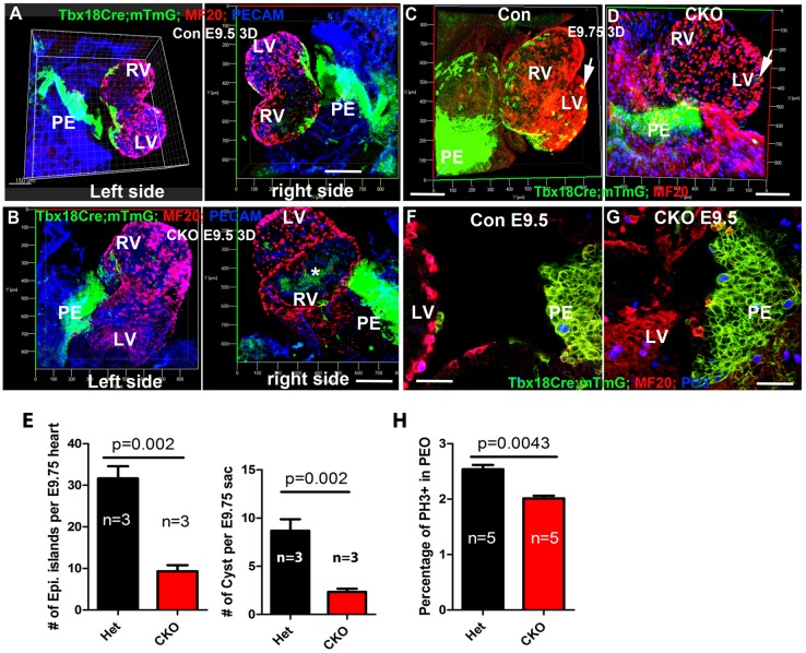
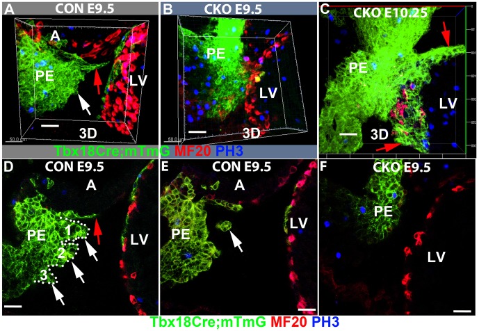
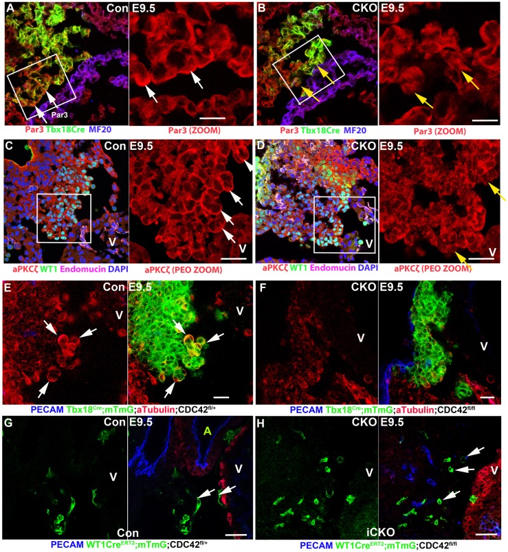
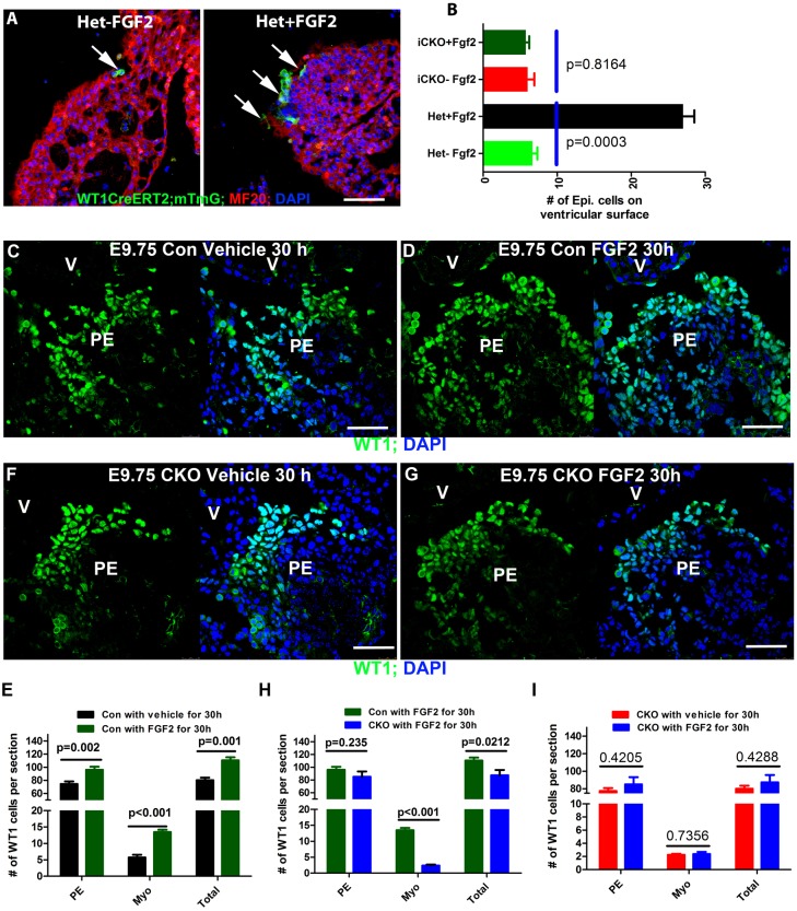
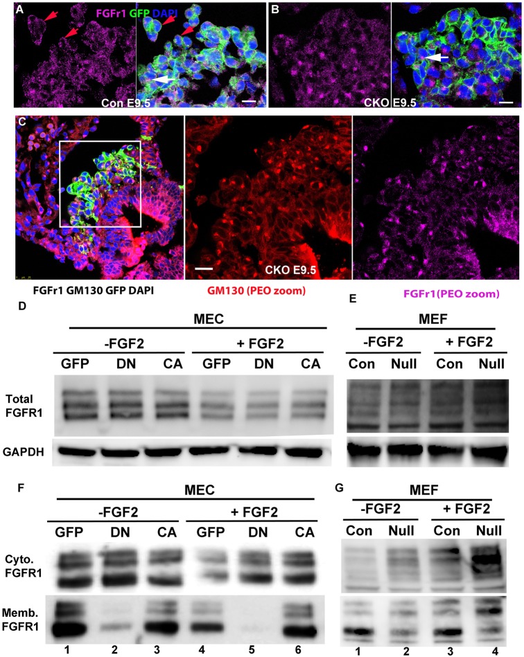
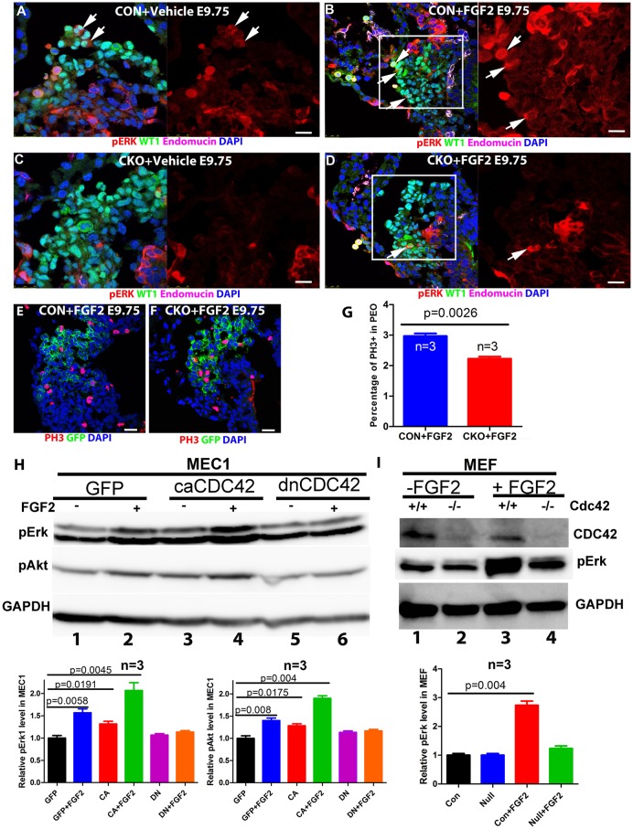
The epicardium contributes to multiple cardiac lineages and is essential for cardiac development and regeneration. However, the mechanism of epicardium formation is unclear. This study aimed to establish the cellular and molecular mechanisms underlying the dissociation of pro-epicardial cells (PECs) from the pro-epicardium (PE) and their subsequent translocation to the heart to form the epicardium. We used lineage tracing, conditional deletion, mosaic analysis and ligand stimulation in mice to determine that both villous protrusions and floating cysts contribute to PEC translocation to myocardium in a CDC42-dependent manner. We resolved a controversy by demonstrating that physical contact of the PE with the myocardium constitutes a third mechanism for PEC translocation to myocardium, and observed a fourth mechanism in which PECs migrate along the surface of the inflow tract to reach the ventricles. Epicardial-specific deletion disrupted epicardium formation, and null PECs proliferated less, lost polarity and failed to form villous protrusions and floating cysts. FGF signaling promotes epicardium formation , and biochemical studies demonstrated that CDC42 is involved in the trafficking of FGF receptors to the cell membrane to regulate epicardium formation.
心外膜对多种心脏谱系有贡献,对心脏发育和再生至关重要。然而,心外膜形成的机制尚不清楚。本研究旨在确定前心外膜细胞(PEC)从前心外膜(PE)解离并随后转移至心脏形成心外膜的细胞和分子机制。我们在小鼠中使用谱系追踪、条件性缺失、镶嵌分析和配体刺激,以确定绒毛状突起和漂浮囊肿均以依赖CDC42的方式促进PEC转移至心肌。我们通过证明PE与心肌的物理接触构成PEC转移至心肌的第三种机制解决了一个争议,并观察到第四种机制,即PEC沿着流入道表面迁移到达心室。心外膜特异性缺失破坏了心外膜形成,空PEC增殖减少、失去极性且无法形成绒毛状突起和漂浮囊肿。FGF信号促进心外膜形成,生化研究表明CDC42参与FGF受体向细胞膜的转运以调节心外膜形成。