Ferraz Raquel, Cunha Clarissa F, Pimentel Maria Inês F, Lyra Marcelo R, Pereira-Da-Silva Tatiana, Schubach Armando O, Da-Cruz Alda Maria, Bertho Alvaro Luiz
Laboratory of Immunoparasitology, Oswaldo Cruz Institute, FIOCRUZ, Rio de Janeiro, RJ, Brazil.
Flow Cytometry Sorting Core Facility, Oswaldo Cruz Institute, FIOCRUZ, Rio de Janeiro, RJ, Brazil.
Parasit Vectors. 2017 May 3;10(1):219. doi: 10.1186/s13071-017-2152-2.
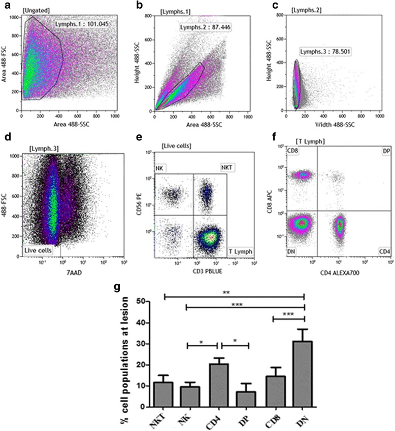
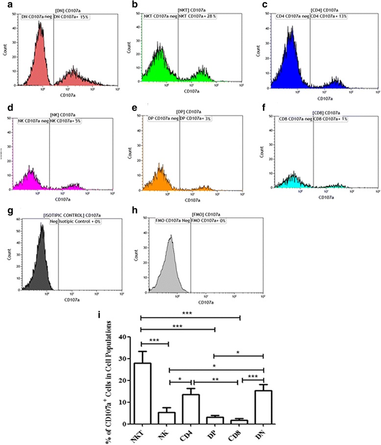
Cutaneous leishmaniasis (CL) is caused by Leishmania (Viannia) braziliensis, which infects dermal macrophages and dendritic cells, causing an intense immune-mediated-tissue inflammation and a skin ulcer with elevated borders that can heal spontaneously or after antimonial therapy. The resolution of lesions depends on an adaptive immune response, and cytotoxic cells seem to have a fundamental role in this process. The aim of this study is to better understand the role of cytotoxicity mediated mechanisms that occur during the immune response in the CL lesion milieu, considering distinct cytotoxic-related CD107a cells, such as CD8, CD4, CD4 CD8 (double-negative, DN) and CD4CD8 (double-positive, DP) T lymphocytes, as well as NK and NKT cells.
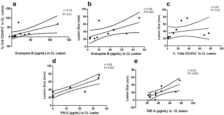
Lesion derived cells were assessed for T cell subpopulations and NK cells, as well as CD107a expression by flow cytometry. In addition, cytometric bead array (CBA) was used to quantify cytokines and granzyme B concentrations in supernatants from macerated lesions.
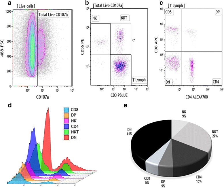
Flow cytometry analyses revealed that NKT cells are the major CD107a-expressing cell population committed to cytotoxicity in CL lesion, although we also observed high frequencies of CD4 and DN T cells expressing CD107a. Analysing the pool of CD107a-cell populations, we found a higher distribution of DN T cells (44%), followed by approximately 25% of NKT cells. Interestingly, NK and CD8 T cells represented only 3 and 4% of the total-CD107a-cell pool, respectively.
The cytotoxicity activity that occurs in the lesion milieu of CL patients seems to be dominated by DN T and NKT cells. These findings suggest the need for a reevaluation of the role of classical-cytotoxic NK and CD8 T cells in the pathogenesis of CL, implicating an important role for other T cell subpopulations.
皮肤利什曼病(CL)由巴西利什曼原虫(维扬亚利什曼原虫)引起,该原虫感染真皮巨噬细胞和树突状细胞,引发强烈的免疫介导组织炎症以及边界隆起的皮肤溃疡,这种溃疡可自发愈合或在锑剂治疗后愈合。病变的消退取决于适应性免疫反应,细胞毒性细胞似乎在这一过程中起关键作用。本研究的目的是更好地理解在CL病变环境中免疫反应期间发生的细胞毒性介导机制的作用,考虑不同的细胞毒性相关CD107a细胞,如CD8、CD4、CD4 CD8(双阴性,DN)和CD4CD8(双阳性,DP)T淋巴细胞,以及NK和NKT细胞。
通过流式细胞术评估病变来源细胞的T细胞亚群、NK细胞以及CD107a表达。此外,采用细胞计数珠阵列(CBA)定量磨碎病变上清液中的细胞因子和颗粒酶B浓度。
流式细胞术分析显示,NKT细胞是CL病变中主要的表达CD107a且具有细胞毒性的细胞群体,尽管我们也观察到表达CD107a的CD4和DN T细胞频率较高。分析CD107a细胞群体库时,我们发现DN T细胞分布更高(44%),其次约为25%的NKT细胞。有趣的是,NK和CD8 T细胞分别仅占总CD107a细胞库的3%和4%。
CL患者病变环境中发生的细胞毒性活性似乎由DN T细胞和NKT细胞主导。这些发现表明需要重新评估经典细胞毒性NK和CD8 T细胞在CL发病机制中的作用,这意味着其他T细胞亚群具有重要作用。