Chandran Sujay, Watkins Johnathan, Abdul-Aziz Amina, Shafat Manar, Calvert Patrick A, Bowles Kristian M, Flather Marcus D, Rushworth Stuart A, Ryding Alisdair D
Norfolk and Norwich University Hospital, Norwich, United Kingdom.
Norwich Medical School, University of East Anglia, Norwich, United Kingdom.
J Am Heart Assoc. 2017 May 3;6(5):e005868. doi: 10.1161/JAHA.117.005868.
Plaque erosion causes 30% of ST-segment elevation myocardial infarctions, but the underlying cause is unknown. Inflammatory infiltrates are less abundant in erosion compared with rupture in autopsy studies. We hypothesized that erosion and rupture are associated with significant differences in intracoronary cytokines in vivo.
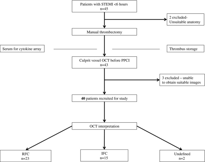
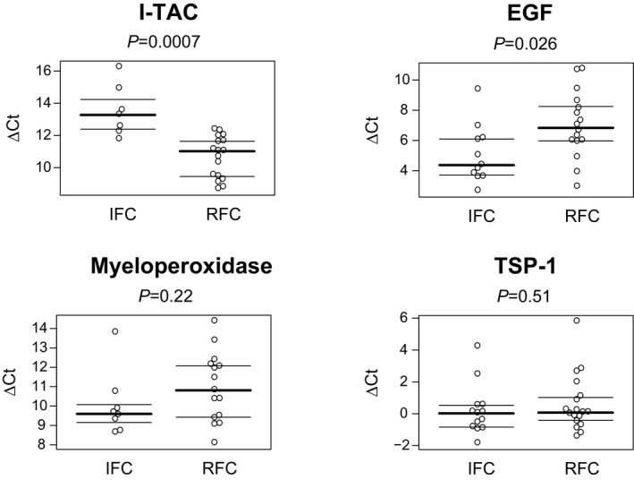
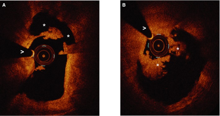
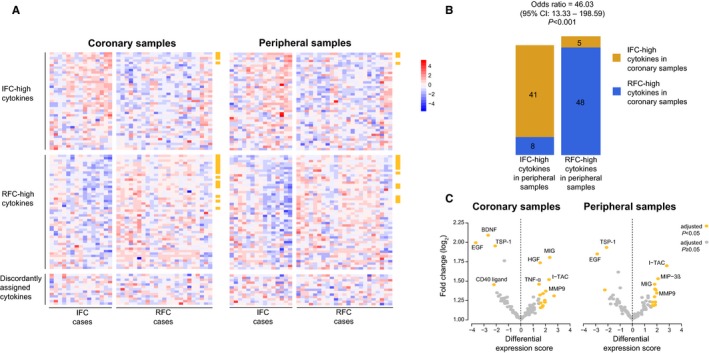
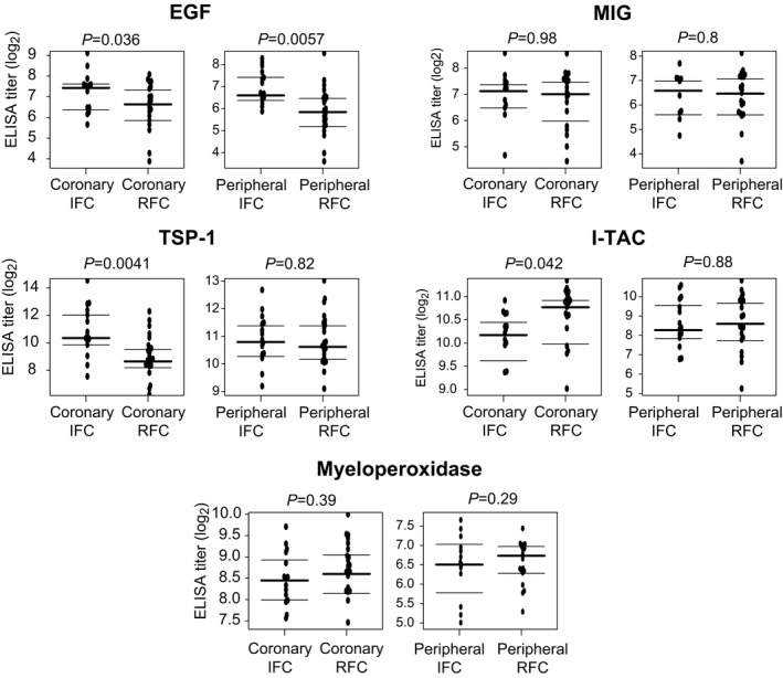
Forty ST-segment elevation myocardial infarction patients with <6 hours of chest pain were classified as ruptured fibrous cap (RFC) or intact fibrous cap (IFC) using optical coherence tomography. Plasma samples from the infarct-related artery and a peripheral artery were analyzed for expression of 102 cytokines using arrays; results were confirmed with ELISA. Thrombectomy samples were analyzed for differential mRNA expression using quantitative real-time polymerase chain reaction. Twenty-three lesions were classified as RFC (58%), 15 as IFC (38%), and 2 were undefined (4%). In addition, 12% (12 of 102) of cytokines were differentially expressed in both coronary and peripheral plasma. I-TAC was preferentially expressed in RFC (significance analysis of microarrays adjusted <0.001; ELISA IFC 10.2 versus RFC 10.8 log pg/mL; =0.042). IFC was associated with preferential expression of epidermal growth factor (significance analysis of microarrays adjusted <0.001; ELISA IFC 7.42 versus RFC 6.63 log pg/mL, =0.036) and thrombospondin 1 (significance analysis of microarrays adjusted =0.03; ELISA IFC 10.4 versus RFC 8.65 log ng/mL, =0.0041). Thrombectomy mRNA showed elevated I-TAC in RFC (=0.0007) epidermal growth factor expression in IFC (=0.0264) but no differences in expression of thrombospondin 1.
These results demonstrate differential intracoronary cytokine expression in RFC and IFC. Elevated thrombospondin 1 and epidermal growth factor may play an etiological role in erosion.
斑块侵蚀导致30%的ST段抬高型心肌梗死,但潜在病因尚不清楚。尸检研究显示,与斑块破裂相比,侵蚀处的炎症浸润较少。我们推测,侵蚀和破裂与体内冠状动脉内细胞因子的显著差异有关。
40例胸痛时间<6小时的ST段抬高型心肌梗死患者,使用光学相干断层扫描将其分为纤维帽破裂(RFC)或纤维帽完整(IFC)。使用阵列分析梗死相关动脉和外周动脉的血浆样本中102种细胞因子的表达;结果用酶联免疫吸附测定(ELISA)进行确认。使用定量实时聚合酶链反应分析血栓切除术样本的差异mRNA表达。23个病变被分类为RFC(58%),15个为IFC(38%),2个未明确(4%)。此外,12%(102种中的12种)的细胞因子在冠状动脉和外周血浆中差异表达。γ-干扰素诱导蛋白10(I-TAC)在RFC中优先表达(微阵列显著性分析校正<0.001;ELISA法测定IFC为10.2,RFC为10.8 log pg/mL;P=0.042)。IFC与表皮生长因子的优先表达相关(微阵列显著性分析校正<0.001;ELISA法测定IFC为7.42,RFC为6.63 log pg/mL,P=0.036)以及血小板反应蛋白1(微阵列显著性分析校正=0.03;ELISA法测定IFC为10.4,RFC为8.65 log ng/mL,P=0.0041)。血栓切除术mRNA显示RFC中I-TAC升高(P=0.0007),IFC中表皮生长因子表达升高(P=0.0264),但血小板反应蛋白1的表达无差异。
这些结果表明RFC和IFC中冠状动脉内细胞因子表达存在差异。血小板反应蛋白1和表皮生长因子升高可能在侵蚀中起病因学作用。