• 文献检索
  • 文档翻译
  • 深度研究
  • 学术资讯
  • Suppr Zotero 插件Zotero 插件
  • 邀请有礼
  • 套餐&价格
  • 历史记录
应用&插件
Suppr Zotero 插件Zotero 插件浏览器插件Mac 客户端Windows 客户端微信小程序
定价
高级版会员购买积分包购买API积分包
服务
文献检索文档翻译深度研究API 文档MCP 服务
关于我们
关于 Suppr公司介绍联系我们用户协议隐私条款
关注我们

Suppr 超能文献

核心技术专利:CN118964589B侵权必究
粤ICP备2023148730 号-1Suppr @ 2026

文献检索

告别复杂PubMed语法,用中文像聊天一样搜索,搜遍4000万医学文献。AI智能推荐,让科研检索更轻松。

立即免费搜索

文件翻译

保留排版,准确专业,支持PDF/Word/PPT等文件格式,支持 12+语言互译。

免费翻译文档

深度研究

AI帮你快速写综述,25分钟生成高质量综述,智能提取关键信息,辅助科研写作。

立即免费体验

与荧光透视引导下经支气管活检相比,支气管内超声联合荧光透视引导下活检获取周围型肺病变样本的系统评价和荟萃分析。

Endobronchial ultrasound plus fluoroscopy-guided biopsy compared to fluoroscopy-guided transbronchial biopsy for obtaining samples of peripheral pulmonary lesions: A systematic review and meta-analysis.

作者信息

Ye Jian, Zhang Ruifeng, Ma Shenglin, Wang Limin, Jin Weizhong

机构信息

Department of Pulmonary Medicine, Hangzhou First People's Hospital, Nanjing Medical University, Hangzhou, China.

Department of Pulmonary Medicine, Sir Run Run Shaw Hospital, Medical School of Zhejiang University, Hangzhou, China.

出版信息

Ann Thorac Med. 2017 Apr-Jun;12(2):114-120. doi: 10.4103/atm.ATM_298_16.

DOI:10.4103/atm.ATM_298_16
PMID:28469722
原文链接:https://pmc.ncbi.nlm.nih.gov/articles/PMC5399685/
Abstract

BACKGROUND

We report a meta-analysis of recent studies comparing the diagnostic yields of endobronchial ultrasonography plus fluoroscopically-guided transbronchial biopsy (EBUS + TBB) with that of conventional fluoroscopically-guided TBB for peripheral pulmonary lesions (PPLs).

METHODS

We searched Medline, the Cochrane Library, PubMed, and Google Scholar through 31 March 2013 using the keywords: lung neoplasm, pulmonary lesions, diagnosis, endobronchial ultrasound, fluoroscopy, and fluoroscopic.

RESULTS

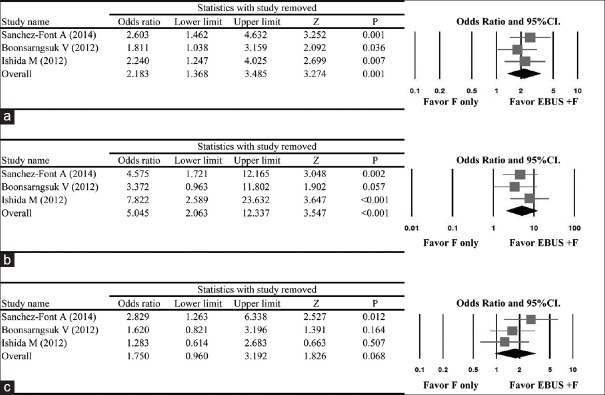
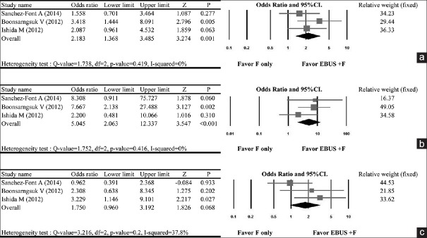
Four studies were included in the study with a total of 461 patients, 222 in the EBUS + TBB group and 239 in the TBB only group. The meta-analysis revealed that the group with EBUS + TBB was more favored in terms of positive diagnostic yield than the group diagnosed with only conventional TBB (odds ratio [OR] = 2.211, 95% confidence interval [CI] = 1.422-3.438, < 0.001). Subgroup analysis based on lesion size found that smaller PPLs had higher accuracy (OR = 4.502, 95% CI = 2.002-10.126, < 0.001) than PPLs of large size (OR = 1.849, 95% CI = 1.033-3.311, = 0.039).

CONCLUSION

Obtaining TBB samples for histopathological diagnosis is enhanced by the addition of EBUS to conventional fluoroscopic guidance; this is, especially important for patients with small peripheral lung lesions who benefit greatly from early diagnosis.

摘要

背景

我们报告一项近期研究的荟萃分析,该分析比较了支气管内超声检查联合荧光镜引导下经支气管活检(EBUS + TBB)与传统荧光镜引导下经支气管活检对周围型肺病变(PPL)的诊断率。

方法

我们使用关键词“肺肿瘤”“肺病变”“诊断”“支气管内超声”“荧光镜检查”和“荧光镜的”,在截至2013年3月31日的Medline、Cochrane图书馆、PubMed和谷歌学术中进行检索。

结果

该研究纳入了四项研究,共461例患者,其中EBUS + TBB组222例,单纯TBB组239例。荟萃分析显示,在阳性诊断率方面,EBUS + TBB组比仅采用传统TBB诊断的组更具优势(优势比[OR] = 2.211,95%置信区间[CI] = 1.422 - 3.438,P < 0.001)。基于病变大小的亚组分析发现,较小的PPL比大尺寸的PPL具有更高的准确性(OR = 4.502,95% CI = 2.002 - 10.126,P < 0.001)(OR = 1.849,95% CI = 1.033 - 3.311,P = 0.039)。

结论

在传统荧光镜引导的基础上增加EBUS可提高获取用于组织病理学诊断的TBB样本的成功率;这对于从小型周围型肺病变中获益于早期诊断的患者尤为重要。

https://cdn.ncbi.nlm.nih.gov/pmc/blobs/bc93/5399685/71f93d1f01bd/ATM-12-114-g004.jpg
https://cdn.ncbi.nlm.nih.gov/pmc/blobs/bc93/5399685/d897627fcb89/ATM-12-114-g001.jpg
https://cdn.ncbi.nlm.nih.gov/pmc/blobs/bc93/5399685/0ca595005666/ATM-12-114-g003.jpg
https://cdn.ncbi.nlm.nih.gov/pmc/blobs/bc93/5399685/71f93d1f01bd/ATM-12-114-g004.jpg
https://cdn.ncbi.nlm.nih.gov/pmc/blobs/bc93/5399685/d897627fcb89/ATM-12-114-g001.jpg
https://cdn.ncbi.nlm.nih.gov/pmc/blobs/bc93/5399685/0ca595005666/ATM-12-114-g003.jpg
https://cdn.ncbi.nlm.nih.gov/pmc/blobs/bc93/5399685/71f93d1f01bd/ATM-12-114-g004.jpg

相似文献

1
Endobronchial ultrasound plus fluoroscopy-guided biopsy compared to fluoroscopy-guided transbronchial biopsy for obtaining samples of peripheral pulmonary lesions: A systematic review and meta-analysis.与荧光透视引导下经支气管活检相比,支气管内超声联合荧光透视引导下活检获取周围型肺病变样本的系统评价和荟萃分析。
Ann Thorac Med. 2017 Apr-Jun;12(2):114-120. doi: 10.4103/atm.ATM_298_16.
2
Efficacy of endobronchial ultrasound-guided transbronchial biopsy without guide sheath for small peripheral pulmonary lesions (≤15 mm): A retrospective cohort study.经支气管超声引导无鞘支气管内活检术用于≤15mm 外周肺部小病变的疗效:一项回顾性队列研究。
Clin Respir J. 2021 Jun;15(6):622-627. doi: 10.1111/crj.13324. Epub 2021 Apr 2.
3
Diagnostic yield of radial probe endobronchial ultrasonography-guided transbronchial biopsy without fluoroscopy in peripheral pulmonary lesions: A systematic review and meta-analysis.径向探头支气管内超声引导经支气管活检术在周围性肺部病变中无透视诊断率的系统评价和荟萃分析。
Thorac Cancer. 2023 Jan;14(2):195-205. doi: 10.1111/1759-7714.14733. Epub 2022 Dec 8.
4
Transbronchial biopsy using endobronchial ultrasonography with a guide sheath increased the diagnostic yield of peripheral pulmonary lesions.使用带有引导鞘的支气管内超声进行经支气管活检可提高周围性肺病变的诊断率。
Intern Med. 2012;51(5):455-60. doi: 10.2169/internalmedicine.51.6358. Epub 2012 Mar 1.
5
Comparison of different transbronchial biopsy sampling techniques for the diagnosis of peripheral pulmonary lesions with radial endobronchial ultrasound-guided bronchoscopy: A prospective study.经支气管镜超声引导下经支气管活检不同采样技术诊断周围型肺病变的比较:一项前瞻性研究。
Respir Investig. 2020 Sep;58(5):381-386. doi: 10.1016/j.resinv.2020.03.004. Epub 2020 Apr 11.
6
Cone-Beam Computed Tomography-Derived Augmented Fluoroscopy Improves the Diagnostic Yield of Endobronchial Ultrasound-Guided Transbronchial Biopsy for Peripheral Pulmonary Lesions.锥形束计算机断层扫描衍生的增强荧光透视术提高了支气管内超声引导下经支气管活检对周围型肺病变的诊断率。
Diagnostics (Basel). 2021 Dec 25;12(1):41. doi: 10.3390/diagnostics12010041.
7
Diagnostic value of the combined use of radial probe endobronchial ultrasound and transbronchial biopsy in lung cancer.径向探头支气管内超声联合经支气管活检对肺癌的诊断价值。
Thorac Cancer. 2020 Jun;11(6):1533-1540. doi: 10.1111/1759-7714.13425. Epub 2020 Apr 16.
8
Endobronchial ultrasound plus fluoroscopy versus fluoroscopy-guided bronchoscopy: a comparison of diagnostic yields in peripheral pulmonary lesions.经支气管超声联合透视与透视引导下经支气管镜检查:对比外周肺部病变的诊断率。
Lung. 2012 Apr;190(2):233-7. doi: 10.1007/s00408-011-9359-3. Epub 2012 Jan 3.
9
Predictive risk factors for pneumothorax after transbronchial biopsy using endobronchial ultrasonography with a guide sheath.经支气管超声引导鞘内活检后发生气胸的预测性风险因素。
BMC Pulm Med. 2021 May 29;21(1):181. doi: 10.1186/s12890-021-01551-1.
10
Using cryoprobes of different sizes combined with cone-beam computed tomography-derived augmented fluoroscopy and endobronchial ultrasound to diagnose peripheral pulmonary lesions: a propensity-matched study.使用不同尺寸的冷冻探头联合锥形束计算机断层扫描衍生的增强荧光透视和支气管内超声诊断周围型肺部病变:一项倾向匹配研究。
Respir Res. 2024 Feb 5;25(1):65. doi: 10.1186/s12931-024-02700-w.

引用本文的文献

1
The incremental contribution of mobile cone-beam computed tomography to the tool-lesion relationship during shape-sensing robotic-assisted bronchoscopy.在形状感知机器人辅助支气管镜检查中,移动锥形束计算机断层扫描对工具-病变关系的增量贡献。
ERJ Open Res. 2024 Jul 22;10(4). doi: 10.1183/23120541.00993-2023. eCollection 2024 Jul.
2
Diagnostic performance of cryobiopsy guided by radial-probe EBUS with a guide sheath for peripheral pulmonary lesions.采用带引导鞘的径向探头超声支气管镜引导下经皮肺穿刺活检术对外周肺部病变的诊断效能。
J Bras Pneumol. 2023 Jan 9;49(1):e20220200. doi: 10.36416/1806-3756/e20220200. eCollection 2023.
3

本文引用的文献

1
Advanced bronchoscopy for the diagnosis of peripheral pulmonary lesions.用于诊断周围型肺部病变的高级支气管镜检查
Respir Investig. 2016 Jul;54(4):224-9. doi: 10.1016/j.resinv.2015.11.008. Epub 2016 Mar 2.
2
Advanced bronchoscopic techniques for the diagnosis of peripheral pulmonary lesions.用于诊断周围型肺部病变的先进支气管镜技术。
Curr Opin Pulm Med. 2016 Jul;22(4):309-18. doi: 10.1097/MCP.0000000000000284.
3
Diagnostic Yield and Complications of Bronchoscopy for Peripheral Lung Lesions. Results of the AQuIRE Registry.
Endobronchial ultrasound-guided transbronchial biopsy with or without a guide sheath for peripheral pulmonary malignancy.
支气管内超声引导下经支气管活检术用于周围型肺恶性肿瘤,可使用或不使用引导鞘。
ERJ Open Res. 2021 Sep 27;7(3). doi: 10.1183/23120541.00267-2021. eCollection 2021 Jul.
4
Diagnostic yield of additional conventional transbronchial lung biopsy following radial endobronchial ultrasound lung biopsy for peripheral pulmonary lesions.经径向超声支气管镜肺活检术(EBUS)检查外周肺部病变后,额外进行常规经支气管肺活检对诊断的影响。
Thorac Cancer. 2020 Jun;11(6):1639-1646. doi: 10.1111/1759-7714.13446. Epub 2020 Apr 27.
5
The value of repeat radial-probe endobronchial ultrasound-guided transbronchial biopsy after initial non-diagnostic results in patients with peripheral pulmonary lesions.在经初始检查结果不明确的外周肺部病变患者中,重复使用径向探头支气管内超声引导经支气管针吸活检的价值。
BMC Pulm Med. 2017 Oct 17;17(1):132. doi: 10.1186/s12890-017-0478-3.
支气管镜检查对周围型肺病变的诊断率及并发症。AQuIRE注册研究结果
Am J Respir Crit Care Med. 2016 Jan 1;193(1):68-77. doi: 10.1164/rccm.201507-1332OC.
4
Endobronchial ultrasound with a guide sheath for small malignant pulmonary nodules: a retrospective comparison between central and peripheral locations.带引导鞘的支气管内超声用于小的恶性肺结节:中央型与外周型的回顾性比较
J Thorac Dis. 2015 Apr;7(4):596-602. doi: 10.3978/j.issn.2072-1439.2015.03.04.
5
Fluoroscopic-Guided Radial Endobronchial Ultrasound Without Guide Sheath For Peripheral Pulmonary Lesions: A Safe And Efficient Combination.荧光透视引导无引导鞘管经支气管径向超声检查用于外周肺部病变:一种安全有效的联合方法。
Arch Bronconeumol. 2015 Jul;51(7):338-43. doi: 10.1016/j.arbres.2014.09.017. Epub 2015 Mar 11.
6
Virtual bronchoscopic navigation for peripheral pulmonary lesions.外周肺病变的虚拟支气管镜导航
Respiration. 2014;88(5):430-40. doi: 10.1159/000367900. Epub 2014 Oct 1.
7
Diagnosis of peripheral pulmonary lesions with radial probe endobronchial ultrasound-guided bronchoscopy.经径向探头支气管内超声引导支气管镜检查诊断周围型肺部病变
Arch Bronconeumol. 2014 Sep;50(9):379-83. doi: 10.1016/j.arbres.2014.02.018. Epub 2014 Apr 18.
8
Radial probe endobronchial ultrasound for peripheral pulmonary lesions. A 5-year institutional experience.径向探头支气管内超声检查用于周围肺部病变。5 年的机构经验。
Ann Am Thorac Soc. 2014 May;11(4):578-82. doi: 10.1513/AnnalsATS.201311-384OC.
9
Endobronchial ultrasound for the diagnosis of peripheral pulmonary lesions. A controlled study with fluoroscopy.支气管内超声用于诊断周围型肺部病变。一项与荧光透视法对照的研究。
Arch Bronconeumol. 2014 May;50(5):166-71. doi: 10.1016/j.arbres.2013.11.019. Epub 2014 Jan 15.
10
Diagnosis of lung nodules with peripheral/radial endobronchial ultrasound-guided transbronchial biopsy.外周/径向支气管内超声引导下经支气管活检诊断肺结节
J Bronchology Interv Pulmonol. 2012 Jan;19(1):5-11. doi: 10.1097/LBR.0b013e31823fcf11.