Department of Radiology, Leiden University Medical Center, Leiden, The Netherlands.
Department of Biomechanical Engineering, Delft University of Technology, Delft, The Netherlands.
Magn Reson Med. 2018 Feb;79(2):1127-1134. doi: 10.1002/mrm.26712. Epub 2017 May 7.
To investigate the feasibility of automatic quantification of bone marrow edema (BME) on MRI of the wrist in patients with early arthritis.
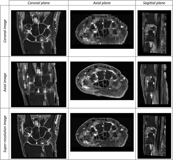
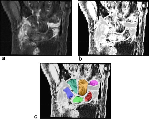
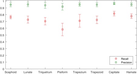
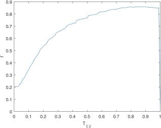
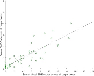
For 485 early arthritis patients (clinically confirmed arthritis of one or more joints, symptoms for less than 2 years), MR scans of the wrist were processed in three automatic stages. First, super-resolution reconstruction was applied to fuse coronal and axial scans into a single high-resolution 3D image. Next, the carpal bones were located and delineated using atlas-based segmentation. Finally, the extent of BME within each bone was quantified by identifying image intensity values characteristic of BME by fuzzy clustering and measuring the fraction of voxels with these characteristic intensities within each bone. Correlation with visual BME scores was assessed through Pearson correlation coefficient.
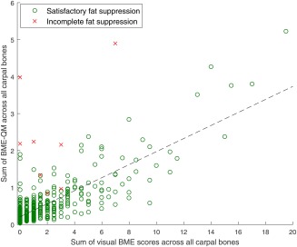
Pearson correlation between quantitative and visual BME scores across 485 patients was r=0.83, P<0.001.
Quantitative measurement of BME on MRI of the wrist has the potential to provide a feasible alternative to visual scoring. Complete automation requires automatic detection and compensation of acquisition artifacts. Magn Reson Med 79:1127-1134, 2018. © 2017 The Authors Magnetic Resonance in Medicine published by Wiley Periodicals, Inc. on behalf of International Society for Magnetic Resonance in Medicine. This is an open access article under the terms of the Creative Commons Attribution-NonCommercial-NoDerivs License, which permits use and distribution in any medium, provided the original work is properly cited, the use is non-commercial and no modifications or adaptations are made.
探究在早期关节炎患者的腕关节 MRI 上自动量化骨髓水肿(BME)的可行性。
对 485 例早期关节炎患者(临床确诊为一个或多个关节的关节炎,症状持续时间<2 年)的腕关节 MRI 进行了三个自动处理阶段。首先,应用超分辨率重建将冠状面和轴向扫描融合成单个高分辨率的 3D 图像。接下来,使用基于图谱的分割定位腕骨并对其进行描绘。最后,通过模糊聚类识别出具有 BME 特征的图像强度值,测量每个骨中具有这些特征强度的体素分数,从而对 BME 的程度进行定量。通过 Pearson 相关系数评估与视觉 BME 评分的相关性。
在 485 例患者中,定量 BME 评分与视觉 BME 评分之间的 Pearson 相关系数为 r=0.83,P<0.001。
腕关节 MRI 上 BME 的定量测量有可能提供一种替代视觉评分的可行方法。完全自动化需要自动检测和补偿采集伪影。磁共振医学 79:1127-1134, 2018。© 2017 作者。磁共振医学由 Wiley 期刊出版公司代表国际磁共振医学学会出版。这是在知识共享署名-非商业性-禁止演绎 4.0 许可协议下的许可,允许在任何媒体中使用、分发,只要原始作品正确引用,使用是非商业性的,并且没有对作品进行修改或改编。