Sung Hsieh Hsiao Hsin, Chung Michael T, Allen Ronald M, Ranganathan Kavitha, Habbouche Joe, Cholok David, Butts Jonathan, Kaura Arminder, Tiruvannamalai-Annamalai Ramkumar, Breuler Chris, Priest Caitlin, Loder Shawn J, Li John, Li Shuli, Stegemann Jan, Kunkel Steven L, Levi Benjamin
Burn/Wound and Regenerative Medicine Laboratory, Department of Surgery, University of Michigan, Ann Arbor, MI, USA.
Experimental Rheumatology, Radboud University Medical Center, Nijmegen, Netherlands.
Front Endocrinol (Lausanne). 2017 Apr 24;8:74. doi: 10.3389/fendo.2017.00074. eCollection 2017.
Heterotopic ossification (HO) occurs in the setting of persistent systemic inflammation. The identification of reliable biomarkers can serve as an early diagnostic tool for HO, especially given the current lack of effective treatment strategies. Although serum biomarkers have great utility, they can be inappropriate or ineffective in traumatic acute injuries and in patients with fibrodysplasia ossificans progressiva (FOP). Therefore, the goal of this study is to profile the cytokines associated with HO using a different non-invasive source of biomarkers.
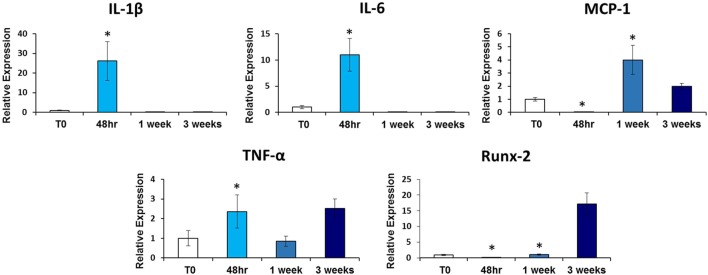
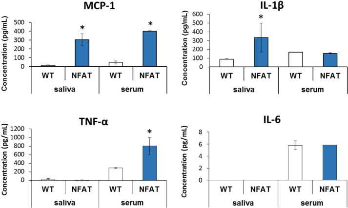
Serum and saliva were collected from a model of trauma-induced HO (tHO) with hind limb Achilles' tenotomy and dorsal burn injury at indicated time points (pre-injury, 48 h, 1 week, and 3 weeks post-injury) and a genetic non-trauma HO model ( ). Samples were analyzed for 27 cytokines using the Bio-Plex assay. Histologic evaluation was performed in mice and at 48 h and 1 week post-injury in burn tenotomy mice. The mRNA expression levels of these cytokines at the tenotomy site were also quantified with quantitative real-time PCR. Pearson correlation coefficient was assessed between saliva and serum.
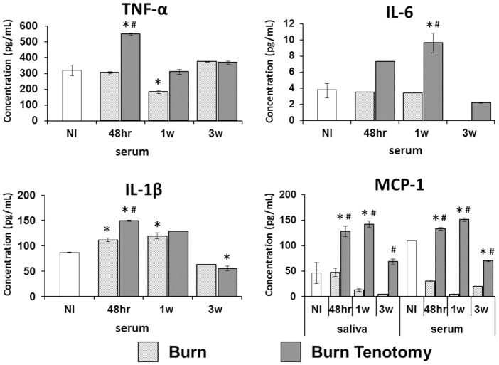
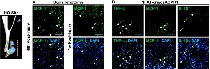
Levels of TNF-α and IL-1β peaked at 48 h and 1 week post-injury in the burn/tenotomy cohort, and these values were significantly higher when compared with both uninjured ( < 0.01, < 0.03) and burn-only mice ( < 0.01, < 0.01). Immunofluorescence staining confirmed enhanced expression of IL-1β, TNF-α, and MCP-1 at the tenotomy site 48 h after injury. Monocyte chemoattractant protein-1 (MCP-1) and VEGF was detected in saliva showing elevated levels at 1 week post-injury in our tHO model when compared with both uninjured ( < 0.001, < 0.01) and burn-only mice ( < 0.005, < 0.01). The Pearson correlation between serum MCP-1 and salivary MCP-1 was statistically significant ( = 0.9686, < 0.001) Similarly, the Pearson correlation between serum VEGF and salivary VEGF was statistically significant ( = 0.9709, < 0.05).
In this preliminary study, we characterized the diagnostic potential of specific salivary cytokines that may serve as biomarkers for an early-stage diagnosis of HO. This study identified two candidate biomarkers for further study and suggests a novel method for diagnosis in the context of current difficult diagnosis and risks of current diagnostic methods in certain patients.
异位骨化(HO)发生于持续性全身炎症的背景下。鉴于目前缺乏有效的治疗策略,识别可靠的生物标志物可作为HO的早期诊断工具。尽管血清生物标志物有很大用途,但在创伤性急性损伤以及进行性骨化性纤维发育不良(FOP)患者中可能不适用或无效。因此,本研究的目的是使用不同的非侵入性生物标志物来源来分析与HO相关的细胞因子。
在指定时间点(损伤前、损伤后48小时、1周和3周)从创伤诱导的HO(tHO)模型(后肢跟腱切断术和背部烧伤损伤)和遗传性非创伤性HO模型中收集血清和唾液样本。使用Bio-Plex检测法分析样本中的27种细胞因子。对小鼠进行组织学评估,并在烧伤切断术小鼠损伤后48小时和1周进行评估。还通过定量实时PCR对切断术部位这些细胞因子的mRNA表达水平进行定量。评估唾液和血清之间的Pearson相关系数。
在烧伤/切断术队列中,TNF-α和IL-1β水平在损伤后48小时和1周达到峰值,与未受伤小鼠(<0.01,<0.03)和仅烧伤小鼠相比,这些值显著更高(<0.01,<0.01)。免疫荧光染色证实损伤后48小时切断术部位IL-1β、TNF-α和MCP-1的表达增强。在我们的tHO模型中,与未受伤小鼠(<0.001,<0.01)和仅烧伤小鼠相比,在唾液中检测到单核细胞趋化蛋白-1(MCP-1)和VEGF,其在损伤后1周水平升高(<0.005,<0.01)。血清MCP-1与唾液MCP-1之间的Pearson相关性具有统计学意义(=0.9686,<0.001)。同样,血清VEGF与唾液VEGF之间的Pearson相关性具有统计学意义(=0.9709,<0.05)。
在这项初步研究中,我们描述了特定唾液细胞因子的诊断潜力,这些细胞因子可能作为HO早期诊断的生物标志物。本研究确定了两种候选生物标志物以供进一步研究,并在当前诊断困难以及某些患者当前诊断方法存在风险的背景下提出了一种新的诊断方法。