Fang Yanan, Sui Rubo
Department of Neurology, the First Affiliated Hospital of Jinzhou Medical University, Jinzhou, China.
Afr J Tradit Complement Altern Med. 2016 Aug 12;13(5):17-24. doi: 10.21010/ajtcam.v13i5.3. eCollection 2016.
Vascular dementia (VD) is the most frequent psychiatric complication of stroke, and is often difficult to treat. Incidence rate of vascular cognition impairment is still 70% after stroke in one year (Sui R et al.2011). Stroke patients with VD suffer from a higher mortality rate and have worse functional outcomes and quality of life. However, despite the extensive literatures on this topic, there is no agreement on the causal mechanisms and effective therapy for VD. The objective of this study is to examine if electroacupuncture at the Wangu acupoint (GB 12), whose position is similar to the cerebellar fastigial nucleus, could reduce inflammatory cytokines in the hippocampus of rats with vascular dementia (VD).
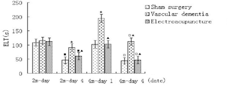
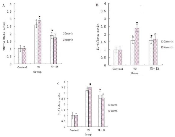
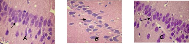
The 54 healthy, male, Sprague-Dawley (SD) rats, 9 months old, and of clean grade (300-450) g, were randomly divided into three groups: sham surgery group, VD group and electro-acupuncture group. The ethology scores of VD rats were evaluated and the mRNA expressions of inflammatory cytokines (TNF-α, IL-6 and IL-1β) in the hippocampus were assessed and the hippocampal tissues were observed by hematoxylin-eosin staining.
Compared with the VD group, in the electroacupuncture group, the rats' learning ability improved significantly and the mRNA expression of TNF-α, IL-6 and IL-1β decreased. Simultaneously, the damage extent of nerve cells in the hippocampal tissues decreased, with their morphology recovered to nearly normal.
Electro-acupuncture at the Wangu acupoint can decrease the levels of inflammatory cytokines in the hippocampus, reduce the damage extent of nerve cells in the hippocampus, and thus provide a new neuroprotective method in VD.
血管性痴呆(VD)是中风最常见的精神并发症,且往往难以治疗。中风一年后血管性认知障碍的发生率仍为70%(隋R等人,2011年)。患有血管性痴呆的中风患者死亡率更高,功能结局和生活质量更差。然而,尽管关于该主题有大量文献,但对于血管性痴呆的病因机制和有效治疗方法尚无共识。本研究的目的是检验位于与小脑顶核位置相似的完骨穴(GB 12)进行电针治疗是否能降低血管性痴呆(VD)大鼠海马中的炎性细胞因子水平。
选取54只9月龄、清洁级(300 - 450)g的健康雄性Sprague-Dawley(SD)大鼠,随机分为三组:假手术组、血管性痴呆组和电针组。评估血管性痴呆大鼠的行为学评分,检测海马中炎性细胞因子(TNF-α、IL-6和IL-1β)的mRNA表达,并通过苏木精-伊红染色观察海马组织。
与血管性痴呆组相比,电针组大鼠的学习能力显著提高且TNF-α、IL-6和IL-1β的mRNA表达降低。同时,海马组织中神经细胞的损伤程度降低,其形态恢复至接近正常。
完骨穴电针治疗可降低海马中炎性细胞因子水平,减轻海马中神经细胞的损伤程度,从而为血管性痴呆提供一种新的神经保护方法。