Huang Lin, Chen Keng, Chen Fu-Chao, Shen Hui-Yong, Ye Ji-Chao, Cai Zhao-Peng, Lin Xi
Department of Orthopedics, Memorial Hospital of Sun Yat-Sen University, Institute of Spinal Cord Injury, Sun Yat-Sen University, Guangzhou, Guangdong Province, 510120, P.R. China.
Department of Ultrasound, Sun Yat-Sen University Cancer Center, State Key Laboratory of Oncology in South China, Guangzhou, Guangdong Province, 510060, P.R. China.
Oncotarget. 2017 Jun 20;8(25):40756-40764. doi: 10.18632/oncotarget.17252.
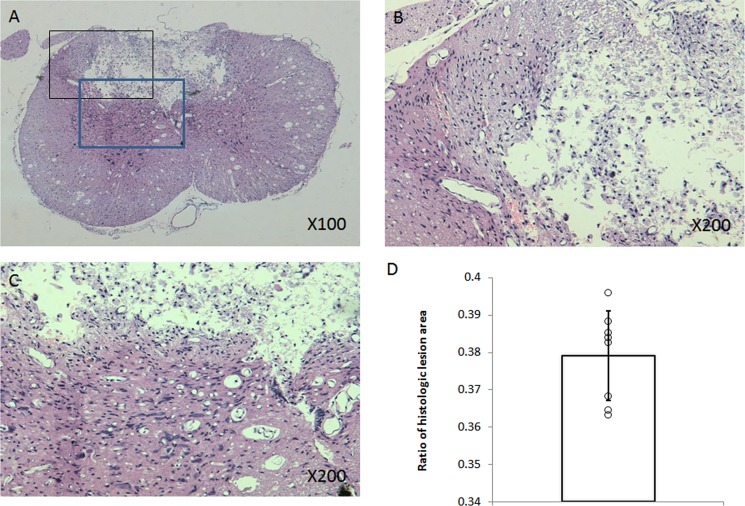
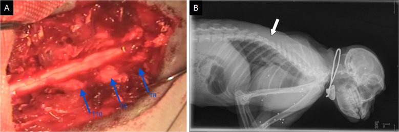
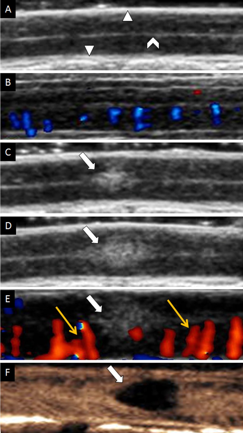
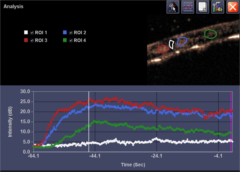
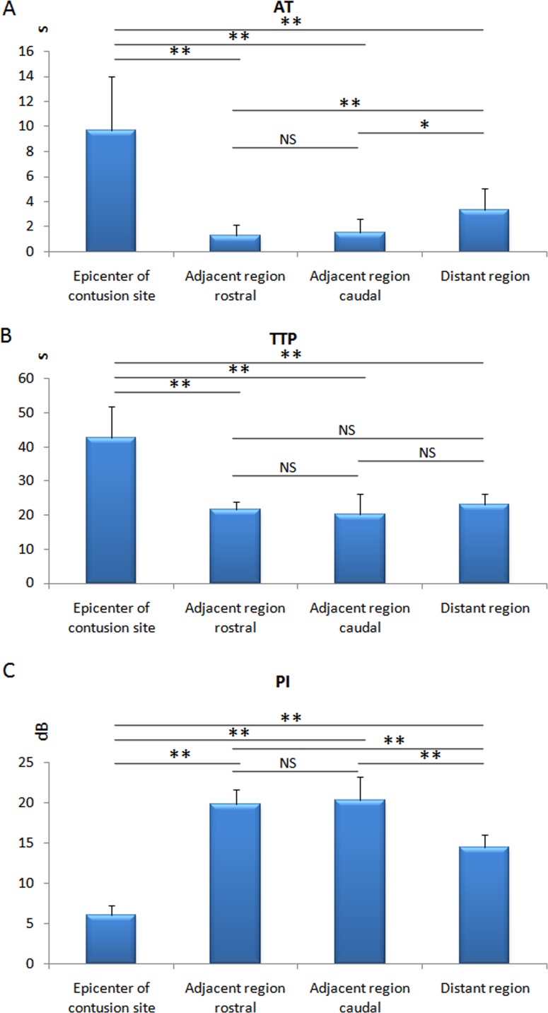
This study tried to quantify spinal cord perfusion by using contrast-enhanced ultrasound (CEUS) in rhesus monkey models with acute spinal cord injury. Acute spinal cord perfusion after injury was detected by CEUS, coupling with conventional ultrasound (US) and Color Doppler US (CDFI). Time-intensity curves and perfusion parameters were obtained by autotracking contrast quantification (ACQ) software in the epicenter and adjacent regions of injury, respectively. Neurological and histological examinations were performed to confirm the severity of injury. US revealed spinal cords were hypoechoic and homogeneous, whereas dura maters, pia maters, and cerebral aqueducts were hyperechoic. After spinal cord contusion, the injured spinal cord was hyperechoic on US, and intramedullary vessels of adjacent region of injury were increased and dilated on CDFI. On CEUS hypoperfusion were found in the epicenter of injury, while hyperperfusion in its adjacent region. Quantitative analysis showed that peak intensity (PI) decreased in epicenters of injury but significantly increased in adjacent regions at all time points (p < 0.05). Functional evaluation demonstrated significant deterioration compared to pre-contusion (p < 0.05). Quantitative analysis with CEUS is a promising method for monitoring perfusion changes of spinal cord injury in overall views and real-time.
本研究试图在恒河猴急性脊髓损伤模型中,通过使用超声造影(CEUS)对脊髓灌注进行量化。通过CEUS检测损伤后的急性脊髓灌注情况,并结合传统超声(US)和彩色多普勒超声(CDFI)。分别通过自动追踪造影剂定量(ACQ)软件在损伤中心和相邻区域获取时间-强度曲线和灌注参数。进行神经学和组织学检查以确认损伤的严重程度。超声显示脊髓呈低回声且均匀,而硬脑膜、软脑膜和脑导水管呈高回声。脊髓挫伤后,损伤的脊髓在超声上呈高回声,损伤相邻区域的脊髓内血管在CDFI上增多且扩张。在CEUS上,发现损伤中心存在低灌注,而其相邻区域存在高灌注。定量分析表明,损伤中心的峰值强度(PI)在所有时间点均降低,但在相邻区域显著增加(p < 0.05)。功能评估显示与挫伤前相比有显著恶化(p < 0.05)。CEUS定量分析是一种在整体视野和实时监测脊髓损伤灌注变化的有前景的方法。