Iwamoto Tsutomu, Nakamura Takashi, Ishikawa Masaki, Yoshizaki Keigo, Sugimoto Asuna, Ida-Yonemochi Hiroko, Ohshima Hayato, Saito Masahiro, Yamada Yoshihiko, Fukumoto Satoshi
Department of Pediatric Dentistry, Institute of Biomedical Sciences, Tokushima University Graduate School, Kuramoto-cho, Tokushima, Japan.
Division of Pediatric Dentistry, Tohoku University Graduate School of Dentistry, Sendai, Miyagi, Japan.
PLoS One. 2017 May 11;12(5):e0177557. doi: 10.1371/journal.pone.0177557. eCollection 2017.
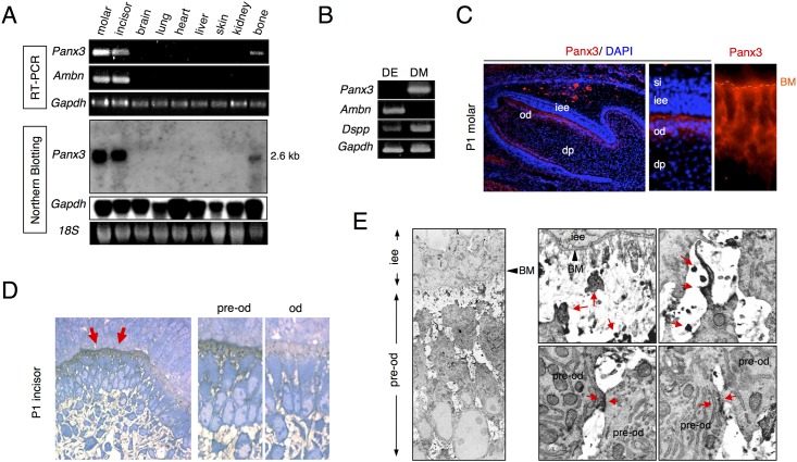
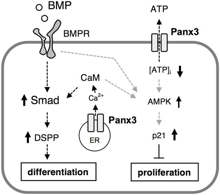
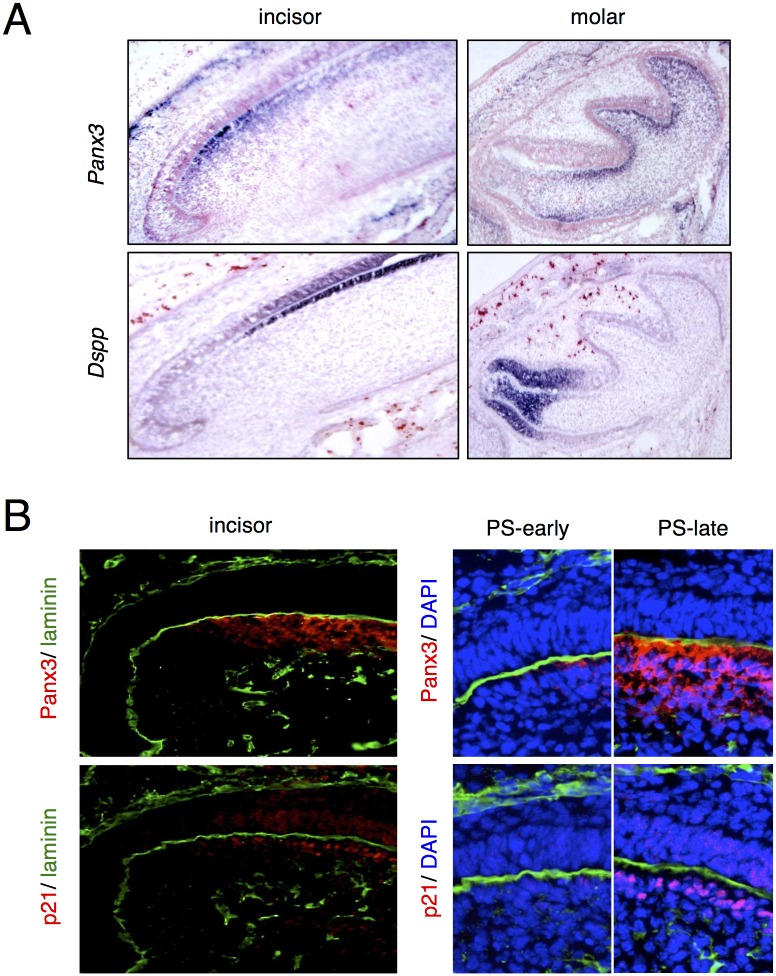
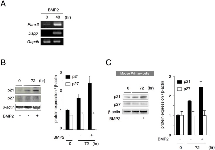
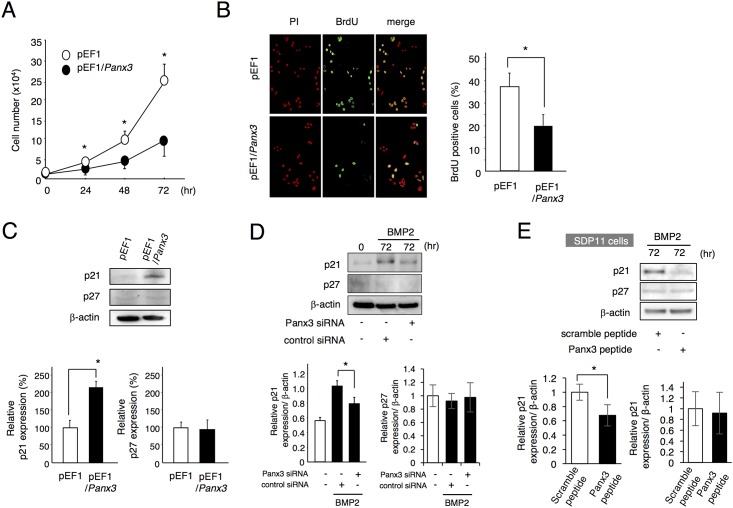
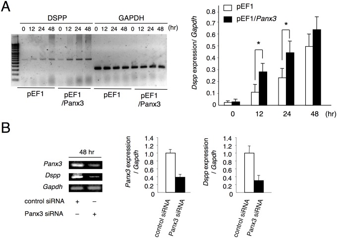
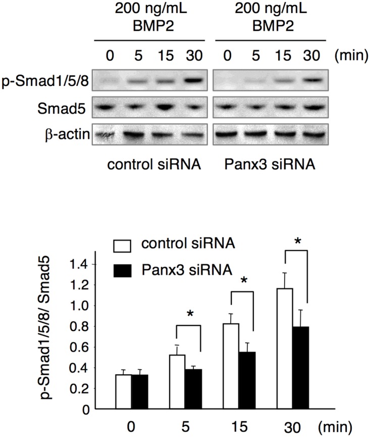
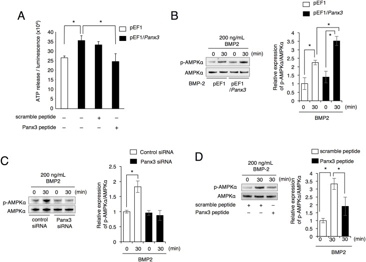
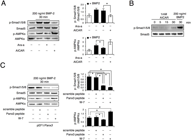
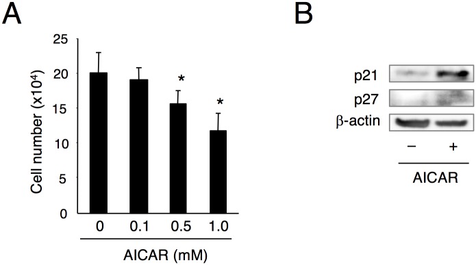
Highly coordinated regulation of cell proliferation and differentiation contributes to the formation of functionally shaped and sized teeth; however, the mechanism underlying the switch from cell cycle exit to cell differentiation during odontogenesis is poorly understood. Recently, we identified pannexin 3 (Panx3) as a member of the pannexin gap junction protein family from tooth germs. The expression of Panx3 was predominately localized in preodontoblasts that arise from dental papilla cells and can differentiate into dentin-secreting odontoblasts. Panx3 also co-localized with p21, a cyclin-dependent kinase inhibitor protein, in preodontoblasts. Panx3 was expressed in primary dental mesenchymal cells and in the mDP dental mesenchymal cell line. Both Panx3 and p21 were induced during the differentiation of mDP cells. Overexpression of Panx3 in mDP cells reduced cell proliferation via up-regulation of p21, but not of p27, and promoted the Bone morphogenetic protein 2 (BMP2)-induced phosphorylation of Smad1/5/8 and the expression of dentin sialophosphoprotein (Dspp), a marker of differentiated odontoblasts. Furthermore, Panx3 released intracellular ATP into the extracellular space through its hemichannel and induced the phosphorylation of AMP-activated protein kinase (AMPK). 5-Aminoimidazole-4-carboxamide-ribonucleoside (AICAR), an activator of AMPK, reduced mDP cell proliferation and induced p21 expression. Conversely, knockdown of endogenous Panx3 by siRNA inhibited AMPK phosphorylation, p21 expression, and the phosphorylation of Smad1/5/8 even in the presence of BMP2. Taken together, our results suggest that Panx3 modulates intracellular ATP levels, resulting in the inhibition of odontoblast proliferation through the AMPK/p21 signaling pathway and promotion of cell differentiation by the BMP/Smad signaling pathway.
细胞增殖与分化的高度协调调控有助于形成功能形状和大小正常的牙齿;然而,牙发生过程中从细胞周期退出到细胞分化转变的潜在机制仍知之甚少。最近,我们从牙胚中鉴定出泛连接蛋白3(Panx3)是泛连接蛋白间隙连接蛋白家族的一员。Panx3的表达主要定位于源自牙乳头细胞并可分化为分泌牙本质的成牙本质细胞的前成牙本质细胞中。Panx3在原代牙间充质细胞和mDP牙间充质细胞系中也有共定位。Panx3和p21(一种细胞周期蛋白依赖性激酶抑制蛋白)在mDP细胞分化过程中均被诱导表达。在mDP细胞中过表达Panx3可通过上调p21而非p27来降低细胞增殖,并促进骨形态发生蛋白2(BMP2)诱导的Smad1/5/8磷酸化以及分化成牙本质细胞标志物牙本质涎磷蛋白(Dspp)的表达。此外,Panx3通过其半通道将细胞内ATP释放到细胞外空间,并诱导AMP活化蛋白激酶(AMPK)的磷酸化。5-氨基咪唑-4-甲酰胺-核苷(AICAR)是一种AMPK激活剂,可降低mDP细胞增殖并诱导p21表达。相反,即使在存在BMP2的情况下,用小干扰RNA(siRNA)敲低内源性Panx3也会抑制AMPK磷酸化、p21表达以及Smad1/5/8的磷酸化。综上所述,我们的结果表明,Panx3调节细胞内ATP水平,通过AMPK/p21信号通路抑制成牙本质细胞增殖,并通过BMP/Smad信号通路促进细胞分化。