Altschwager Pablo, Moskowitz Anne, Fulton Anne B, Hansen Ronald M
Department of Ophthalmology, Boston Children's Hospital and Harvard Medical School, Boston, Massachusetts, United States 2Departamento de Oftalmologia, Escuela de Medicina, Pontificia Universidad Catolica de Chile, Santiago, Chile.
Department of Ophthalmology, Boston Children's Hospital and Harvard Medical School, Boston, Massachusetts, United States.
Invest Ophthalmol Vis Sci. 2017 May 1;58(5):2603-2608. doi: 10.1167/iovs.17-21587.
The purpose of this study is to assess cone-mediated central retinal function in children with a history of preterm birth, including subjects with and without retinopathy of prematurity (ROP). The multifocal electroretinogram (mfERG) records activity of the postreceptor retinal circuitry.
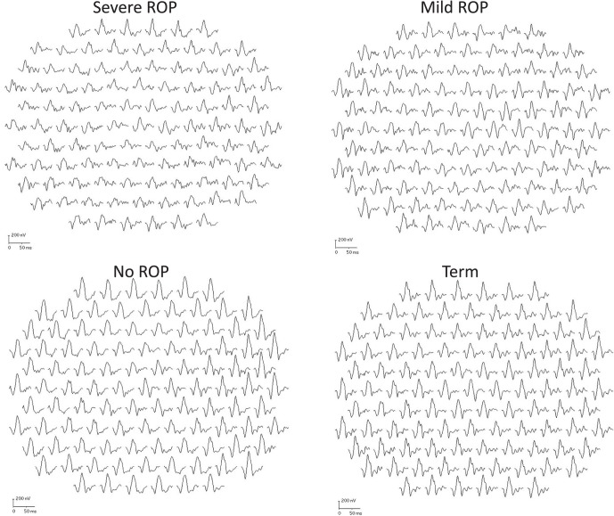
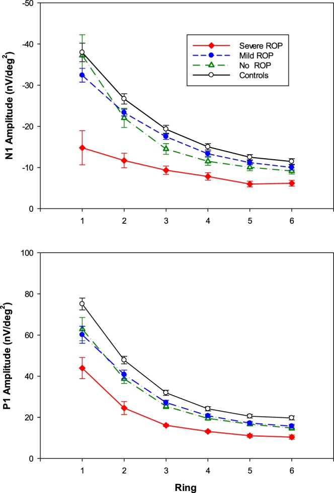
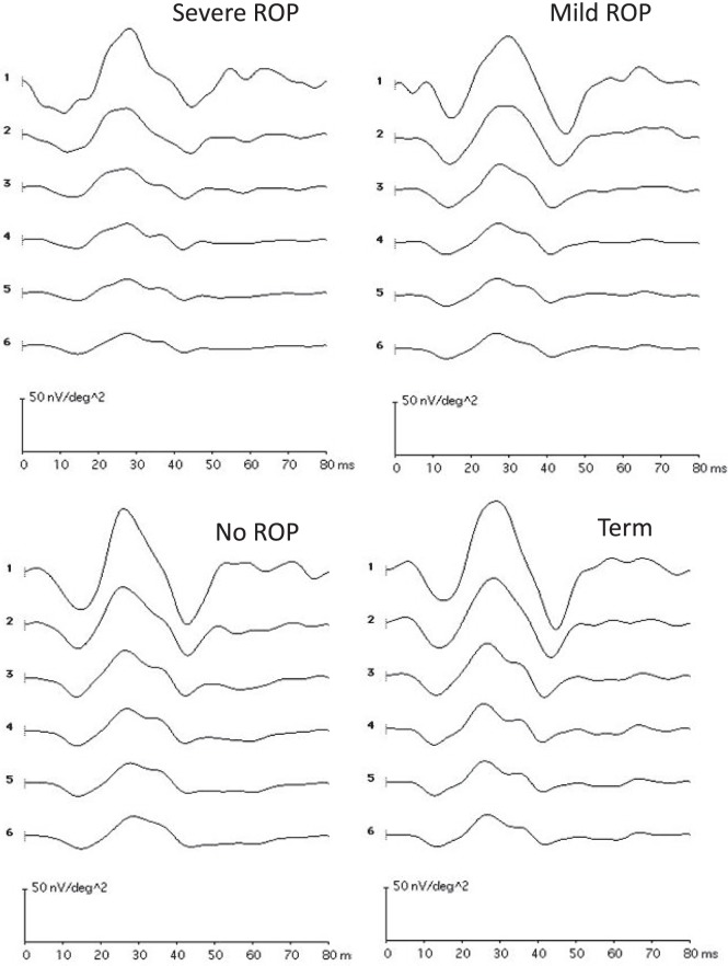
mfERG responses were recorded to an array of 103 hexagonal elements that subtended 43° around a central fixation target. The amplitude and latency of the first negative (N1) and first positive (P1) response were evaluated in six concentric rings centered on the fovea. Responses were recorded from 40 subjects with a history of preterm birth (severe ROP, mild ROP, no ROP) and 19 term-born control subjects.
The amplitude of N1 and P1 varied significantly with eccentricity and ROP severity. For all four groups, these amplitudes were largest in the center and decreased with eccentricity. At all eccentricities, N1 amplitude was significantly smaller in severe ROP and did not differ significantly among the other three groups (mild ROP, no ROP, term-born controls). P1 amplitude in all preterm groups was significantly smaller than in controls; P1 amplitude was similar in no ROP and mild ROP and significantly smaller in severe ROP.
These results provide evidence that premature birth alone affects cone-mediated central retinal function and that the magnitude of the effect varies with severity of the antecedent ROP. The lack of difference in mfERG amplitude between the mild and no ROP groups is evidence that the effect of ROP on the neurosensory retina may not depend solely on appearance of abnormal retinal vasculature.
本研究旨在评估有早产史儿童的视锥细胞介导的视网膜中央功能,包括患有和未患有早产儿视网膜病变(ROP)的受试者。多焦视网膜电图(mfERG)记录感受器后视网膜回路的活动。
mfERG反应记录到围绕中央注视目标的103个六边形单元阵列上。在以中央凹为中心的六个同心环中评估第一个负向(N1)和第一个正向(P1)反应的振幅和潜伏期。记录了40名有早产史的受试者(重度ROP、轻度ROP、无ROP)和19名足月儿对照受试者的反应。
N1和P1的振幅随离心率和ROP严重程度显著变化。对于所有四组,这些振幅在中心最大,并随离心率降低。在所有离心率下,重度ROP组的N1振幅明显较小,而其他三组(轻度ROP、无ROP、足月儿对照)之间无显著差异。所有早产组的P1振幅均显著小于对照组;无ROP组和轻度ROP组的P1振幅相似,重度ROP组明显较小。
这些结果提供了证据,表明仅早产就会影响视锥细胞介导的视网膜中央功能,且影响程度随先前ROP的严重程度而变化。轻度ROP组和无ROP组之间mfERG振幅无差异,这证明ROP对神经感觉视网膜的影响可能不仅仅取决于异常视网膜血管的出现。