• 文献检索
  • 文档翻译
  • 深度研究
  • 学术资讯
  • Suppr Zotero 插件Zotero 插件
  • 邀请有礼
  • 套餐&价格
  • 历史记录
应用&插件
Suppr Zotero 插件Zotero 插件浏览器插件Mac 客户端Windows 客户端微信小程序
定价
高级版会员购买积分包购买API积分包
服务
文献检索文档翻译深度研究API 文档MCP 服务
关于我们
关于 Suppr公司介绍联系我们用户协议隐私条款
关注我们

Suppr 超能文献

核心技术专利:CN118964589B侵权必究
粤ICP备2023148730 号-1Suppr @ 2026

文献检索

告别复杂PubMed语法,用中文像聊天一样搜索,搜遍4000万医学文献。AI智能推荐,让科研检索更轻松。

立即免费搜索

文件翻译

保留排版,准确专业,支持PDF/Word/PPT等文件格式,支持 12+语言互译。

免费翻译文档

深度研究

AI帮你快速写综述,25分钟生成高质量综述,智能提取关键信息,辅助科研写作。

立即免费体验

骨间后神经综合征的超声检查结果

Ultrasonographic findings of posterior interosseous nerve syndrome.

作者信息

Kim Youdong, Ha Doo Hoe, Lee Sang Min

机构信息

Department of Radiology, CHA Bundang Medical Center, CHA University, Seongnam, Korea.

出版信息

Ultrasonography. 2017 Oct;36(4):363-369. doi: 10.14366/usg.17007. Epub 2017 Apr 5.

DOI:10.14366/usg.17007
PMID:28494524
原文链接:https://pmc.ncbi.nlm.nih.gov/articles/PMC5621806/
Abstract

PURPOSE

The purpose of this study was to evaluate the ultrasonographic findings associated with posterior interosseous nerve (PIN) syndrome.

METHODS

Approval from the Institutional Review Board was obtained. A retrospective review of 908 patients' sonographic images of the upper extremity from January 2001 to October 2010 revealed 10 patients suspicious for a PIN abnormality (7 male and 3 female patients; mean age of 51.8±13.1 years; age range, 32 to 79 years). The ultrasonographic findings of PIN syndrome, including changes in the PIN and adjacent secondary changes, were evaluated. The anteroposterior diameter of the pathologic PIN was measured in eight patients and the anteroposterior diameter of the contralateral asymptomatic PIN was measured in six patients, all at the level immediately proximal to the proximal supinator border. The size of the pathologic nerves and contralateral asymptomatic nerves was compared using the Mann-Whitney U test.

RESULTS

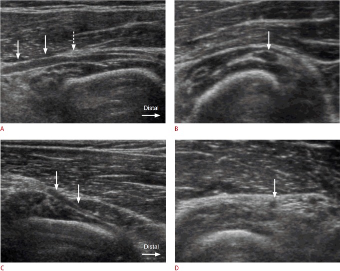
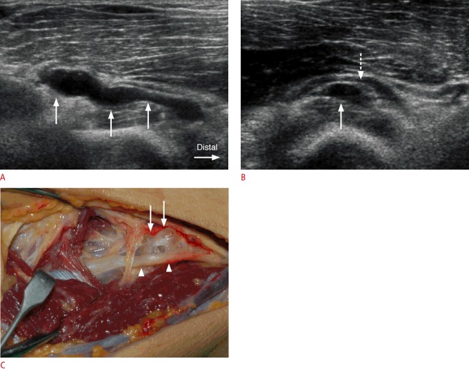
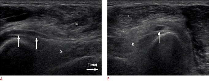
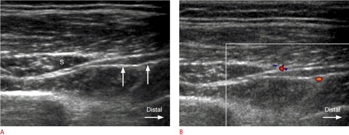
Swelling of the PIN proximal to the supinator canal by compression at the arcade of Fröhse was observed in four cases. Swelling of the PIN distal to the supinator canal was observed in one case. Loss of the perineural fat plane in the supinator canal was observed in one case. Four soft tissue masses were noted. Secondary denervation atrophy of the supinator and extensor muscles was observed in two cases. The mean anteroposterior diameter of the pathologic nerves (n=8, 1.79±0.43 mm) was significantly larger than that of the contralateral asymptomatic nerves (n=6, 1.02±0.22 mm) (P=0.003).

CONCLUSION

Ultrasonography provides high-resolution images of the PIN and helps to diagnose PIN syndrome through visualization of its various causes and adjacent secondary changes.

摘要

目的

本研究旨在评估与骨间后神经(PIN)综合征相关的超声检查结果。

方法

获得了机构审查委员会的批准。对2001年1月至2010年10月期间908例患者上肢的超声图像进行回顾性分析,发现10例患者疑似PIN异常(7例男性和3例女性患者;平均年龄51.8±13.1岁;年龄范围32至79岁)。评估了PIN综合征的超声检查结果,包括PIN的变化和相邻的继发性变化。在8例患者中测量了病理性PIN的前后径,在6例患者中测量了对侧无症状PIN的前后径,均在旋后肌近端边界近端紧邻的水平进行测量。使用Mann-Whitney U检验比较病理性神经和对侧无症状神经的大小。

结果

4例观察到在弗罗伊泽弓处受压导致旋后肌管近端的PIN肿胀。1例观察到旋后肌管远端的PIN肿胀。1例观察到旋后肌管内神经周围脂肪平面消失。发现4个软组织肿块。2例观察到旋后肌和伸肌的继发性失神经萎缩。病理性神经的平均前后径(n = 8,1.79±0.43 mm)明显大于对侧无症状神经的平均前后径(n = 6,1.02±0.22 mm)(P = 0.003)。

结论

超声检查可提供PIN的高分辨率图像,并通过可视化其各种病因和相邻的继发性变化有助于诊断PIN综合征。

https://cdn.ncbi.nlm.nih.gov/pmc/blobs/e10b/5621806/0f7fce8d0c53/usg-17007-f4.jpg
https://cdn.ncbi.nlm.nih.gov/pmc/blobs/e10b/5621806/583623ea4a99/usg-17007-f1.jpg
https://cdn.ncbi.nlm.nih.gov/pmc/blobs/e10b/5621806/b7f5f4c48c6f/usg-17007-f2.jpg
https://cdn.ncbi.nlm.nih.gov/pmc/blobs/e10b/5621806/85dd0667a175/usg-17007-f3.jpg
https://cdn.ncbi.nlm.nih.gov/pmc/blobs/e10b/5621806/0f7fce8d0c53/usg-17007-f4.jpg

相似文献

1
Ultrasonographic findings of posterior interosseous nerve syndrome.骨间后神经综合征的超声检查结果
Ultrasonography. 2017 Oct;36(4):363-369. doi: 10.14366/usg.17007. Epub 2017 Apr 5.
2
Cross-sectional sonographic assessment of the posterior interosseous nerve.骨间后神经的横断面超声评估
Surg Radiol Anat. 2015 Dec;37(10):1155-60. doi: 10.1007/s00276-015-1487-7. Epub 2015 May 13.
3
High resolution ultrasound in posterior interosseous nerve syndrome.高频超声在后骨间神经综合征中的应用。
Muscle Nerve. 2014 Jan;49(1):35-9. doi: 10.1002/mus.23867. Epub 2013 Sep 20.
4
Anatomical Study of Innervation of the Supinator Muscle to Reinnervate the Posterior Interosseous Nerve.对旋后肌神经支配进行再支配骨间后神经的解剖学研究。
Rev Bras Ortop (Sao Paulo). 2019 May;54(3):253-260. doi: 10.1055/s-0039-1692459. Epub 2019 Jun 27.
5
Posterior interosseous nerve of the elbow at the arcade of Frohse: Ultrasound appearance in asymptomatic subjects.肘后窝的桡神经弓:无症状受试者的超声表现。
Diagn Interv Imaging. 2019 Sep;100(9):521-525. doi: 10.1016/j.diii.2019.03.007. Epub 2019 Mar 29.
6
A study of the posterior interosseous nerve (PIN) and the radial tunnel in 30 Thai cadavers.一项对30具泰国尸体的骨间后神经(PIN)和桡管的研究。
J Hand Surg Am. 1993 Jan;18(1):107-12. doi: 10.1016/0363-5023(93)90253-Y.
7
Posterior interosseous nerve of the elbow: normal appearances simulating entrapment.肘部骨间后神经:正常表现酷似卡压。
J Ultrasound Med. 2010 May;29(5):691-6. doi: 10.7863/jum.2010.29.5.691.
8
Morphology of the Posterior Interosseous Nerve with Regard to Entrapment Syndrome.关于卡压综合征的骨间后神经形态学
Indian J Orthop. 2020 Mar 14;54(Suppl 1):188-192. doi: 10.1007/s43465-020-00084-9. eCollection 2020 Sep.
9
Posterior interosseous nerve discontinuity due to compression by lipoma: report of 2 cases.由于脂肪瘤压迫导致的骨间后神经连续性中断:2 例报告。
J Neurosurg. 2017 May;126(5):1698-1701. doi: 10.3171/2016.2.JNS152810. Epub 2016 May 13.
10
Quantitative anatomical study of the posterior interosseous nerve.骨间后神经的定量解剖学研究
Am J Orthop (Belle Mead NJ). 2000 Sep;29(9):702-4.

引用本文的文献

1
Imaging of elbow entrapment neuropathies.肘部卡压性神经病的影像学表现
Insights Imaging. 2025 Jan 29;16(1):24. doi: 10.1186/s13244-025-01901-1.
2
Posterior interosseous nerve lesion due to lipoma. Review of the literature and rare case presentation.因脂肪瘤导致的骨间后神经损伤。文献回顾与罕见病例报告。
Radiol Oncol. 2024 Oct 4;58(4):480-485. doi: 10.2478/raon-2024-0041. eCollection 2024 Dec 1.
3
Imaging characteristics of posterior interosseous nerve syndrome.骨间后神经综合征的影像学特征。

本文引用的文献

1
High resolution ultrasound in posterior interosseous nerve syndrome.高频超声在后骨间神经综合征中的应用。
Muscle Nerve. 2014 Jan;49(1):35-9. doi: 10.1002/mus.23867. Epub 2013 Sep 20.
2
Nerve entrapment syndromes of the elbow, forearm, and wrist.肘部、前臂和腕部的神经卡压综合征。
AJR Am J Roentgenol. 2010 Sep;195(3):585-94. doi: 10.2214/AJR.10.4817.
3
Entrapment of the posterior interosseous nerve at the arcade of Frohse with sonographic, magnetic resonance imaging, and intraoperative confirmation.超声、磁共振成像及术中证实的弗罗瑟弓处骨间后神经卡压
Radiol Case Rep. 2024 Mar 14;19(6):2196-2201. doi: 10.1016/j.radcr.2024.02.090. eCollection 2024 Jun.
4
Role of high-resolution ultrasound and magnetic resonance neurography in the evaluation of peripheral nerves in the upper extremity.高分辨率超声和磁共振神经造影在上肢周围神经评估中的作用。
J Ultrason. 2023 Nov 23;23(95):e313-e327. doi: 10.15557/jou.2023.0037. eCollection 2023 Oct.
5
Treatment of posterior interosseous nerve entrapment syndrome with ultrasound-guided hydrodissection: A case report.超声引导下水分离治疗骨间后神经卡压综合征:一例报告
World J Clin Cases. 2023 Sep 26;11(27):6624-6630. doi: 10.12998/wjcc.v11.i27.6624.
6
An unusual cause of posterior interosseous nerve palsy and contribution of ultrasonography to electromyography in a patient with neurofibroma.一名患有神经纤维瘤患者的骨间后神经麻痹的罕见病因及超声检查对肌电图的辅助作用
Turk J Phys Med Rehabil. 2022 Apr 26;69(3):380-384. doi: 10.5606/tftrd.2023.9881. eCollection 2023 Sep.
7
Orthopedic Management of Radial Tunnel Syndrome: A Diagnostic and Treatment Dilemma.桡骨管综合征的骨科治疗:诊断与治疗难题
Eurasian J Med. 2023 Feb;55(1):59-63. doi: 10.5152/eurasianjmed.2023.22274.
8
Radial nerve compression: anatomical perspective and clinical consequences.桡神经压迫:解剖学视角与临床后果。
Neurosurg Rev. 2023 Feb 13;46(1):53. doi: 10.1007/s10143-023-01944-2.
9
Multihit Injury of the Radial Nerve in a 62-year-old Woman: A Case Report.一名62岁女性桡神经的多发伤:病例报告
Plast Reconstr Surg Glob Open. 2022 Jun 20;10(6):e4414. doi: 10.1097/GOX.0000000000004414. eCollection 2022 Jun.
10
Posterior Interosseous Nerve Graft: Utilizing External Landmarks and Anthropometric Ratios to Predict Available Length for Digital Nerve Reconstruction in a Cadaveric Study.骨间后神经移植:在一项尸体研究中利用外部标志和人体测量比例预测用于指神经重建的可用长度
Plast Surg (Oakv). 2022 May;30(2):130-135. doi: 10.1177/2292550321995731. Epub 2021 May 12.
J Ultrasound Med. 2009 Jun;28(6):807-12. doi: 10.7863/jum.2009.28.6.807.
4
Frohse's arcade is not the exclusive compression site of the radial nerve in its tunnel.弗罗泽氏弓并非桡神经在其隧道内的唯一卡压部位。
Orthop Traumatol Surg Res. 2009 Apr;95(2):114-8. doi: 10.1016/j.otsr.2008.11.001. Epub 2009 Mar 17.
5
Anatomic landmarks for the radial tunnel.桡骨管的解剖标志。
Eplasty. 2008 Jun 22;8:e37.
6
Ultrasonographic findings in isolated neuritis of the posterior interosseous nerve: comparison with normal findings.
J Ultrasound Med. 2007 May;26(5):683-7. doi: 10.7863/jum.2007.26.5.683.
7
The two locations of ganglions causing radial nerve palsy.导致桡神经麻痹的腱鞘囊肿的两个部位。
J Hand Surg Eur Vol. 2007 Jun;32(3):341-5. doi: 10.1016/J.JHSB.2006.09.014.
8
Incidence of common compressive neuropathies in primary care.基层医疗中常见压迫性神经病变的发病率。
J Neurol Neurosurg Psychiatry. 2006 Feb;77(2):263-5. doi: 10.1136/jnnp.2005.066696.
9
Mononeuropathies of the radial nerve: clinical and neurographic findings in 91 consecutive cases.桡神经单神经病:91例连续病例的临床及神经电图检查结果
J Electromyogr Kinesiol. 2005 Aug;15(4):377-83. doi: 10.1016/j.jelekin.2005.01.003.
10
Sonography of entrapment neuropathies in the upper limb (wrist excluded).上肢(不包括腕部)卡压性神经病的超声检查
J Clin Ultrasound. 2004 Nov-Dec;32(9):438-50. doi: 10.1002/jcu.20067.