Sameni Mansoureh, Cavallo-Medved Dora, Franco Omar E, Chalasani Anita, Ji Kyungmin, Aggarwal Neha, Anbalagan Arulselvi, Chen Xuequn, Mattingly Raymond R, Hayward Simon W, Sloane Bonnie F
Department of Pharmacology, Wayne State University School of Medicine, Detroit, MI, 48201, USA.
Department of Biological Sciences, University of Windsor, Windsor, ON, N9B 3P4, Canada.
Breast Cancer Res. 2017 May 15;19(1):56. doi: 10.1186/s13058-017-0847-0.
The breast tumor microenvironment regulates progression of ductal carcinoma in situ (DCIS) to invasive ductal carcinoma (IDC). However, it is unclear how interactions between breast epithelial and stromal cells can drive this progression and whether there are reliable microenvironmental biomarkers to predict transition of DCIS to IDC.
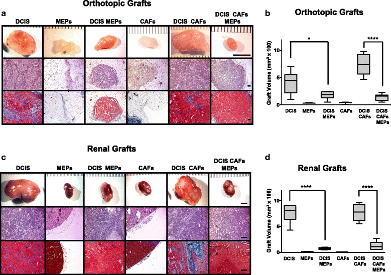
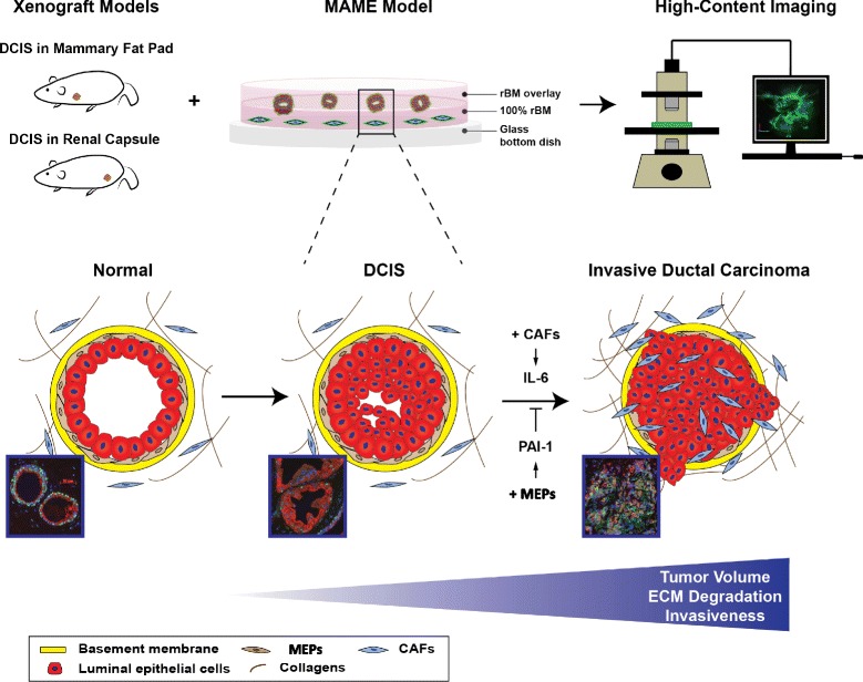
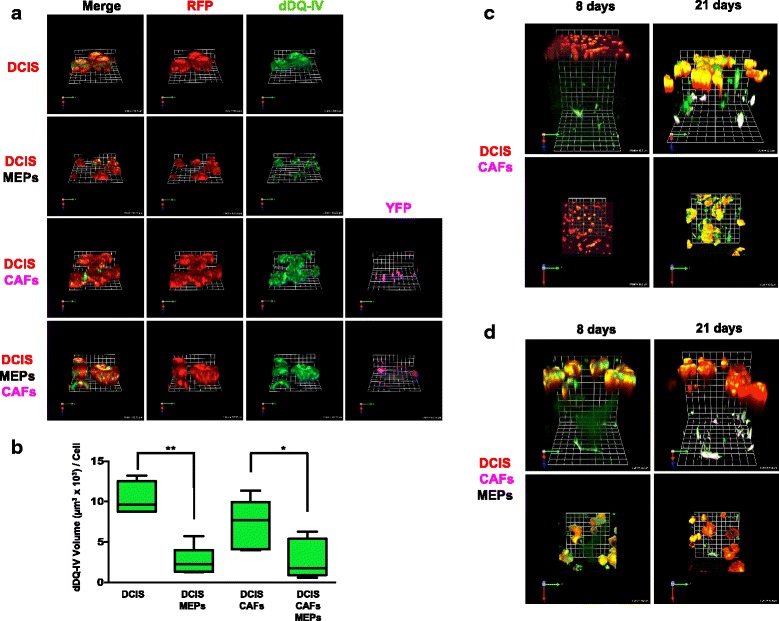
We used xenograft mouse models and a 3D pathomimetic model termed mammary architecture and microenvironment engineering (MAME) to study the interplay between human breast myoepithelial cells (MEPs) and cancer-associated fibroblasts (CAFs) on DCIS progression.
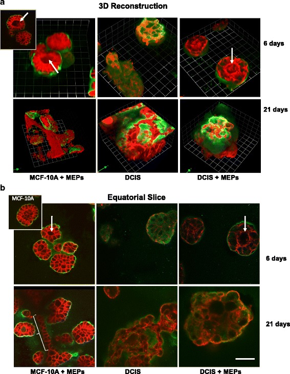
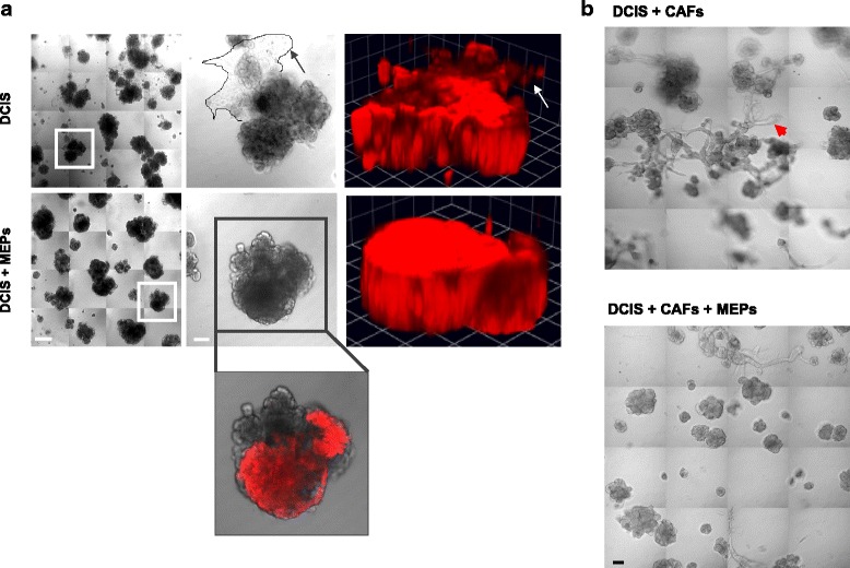
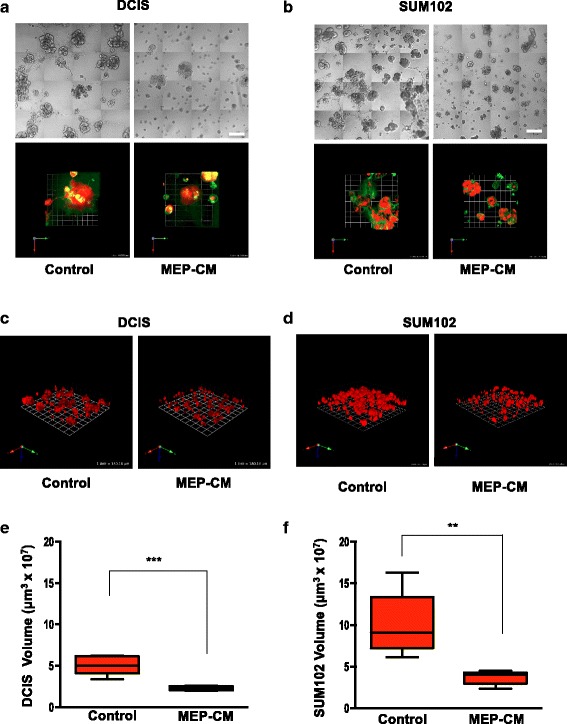
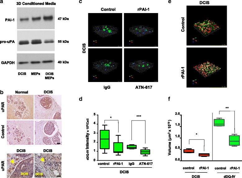
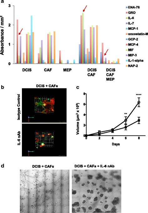
Our results show that MEPs suppress tumor formation by DCIS cells in vivo even in the presence of CAFs. In the in vitro MAME model, MEPs reduce the size of 3D DCIS structures and their degradation of extracellular matrix. We further show that the tumor-suppressive effects of MEPs on DCIS are linked to inhibition of urokinase plasminogen activator (uPA)/urokinase plasminogen activator receptor (uPAR)-mediated proteolysis by plasminogen activator inhibitor 1 (PAI-1) and that they can lessen the tumor-promoting effects of CAFs by attenuating interleukin 6 (IL-6) signaling pathways.
Our studies using MAME are, to our knowledge, the first to demonstrate a divergent interplay between MEPs and CAFs within the DCIS tumor microenvironment. We show that the tumor-suppressive actions of MEPs are mediated by PAI-1, uPA and its receptor, uPAR, and are sustained even in the presence of the CAFs, which themselves enhance DCIS tumorigenesis via IL-6 signaling. Identifying tumor microenvironmental regulators of DCIS progression will be critical for defining a robust and predictive molecular signature for clinical use.
乳腺肿瘤微环境调节导管原位癌(DCIS)向浸润性导管癌(IDC)的进展。然而,尚不清楚乳腺上皮细胞与基质细胞之间的相互作用如何驱动这一进展,以及是否存在可靠的微环境生物标志物来预测DCIS向IDC的转变。
我们使用异种移植小鼠模型和一种称为乳腺结构与微环境工程(MAME)的三维病理模拟模型,研究人乳腺肌上皮细胞(MEP)与癌症相关成纤维细胞(CAF)在DCIS进展中的相互作用。
我们的结果表明,即使在存在CAF的情况下,MEP在体内也能抑制DCIS细胞形成肿瘤。在体外MAME模型中,MEP减小了三维DCIS结构的大小及其对细胞外基质的降解。我们进一步表明,MEP对DCIS的肿瘤抑制作用与纤溶酶原激活物抑制剂1(PAI-1)抑制尿激酶型纤溶酶原激活物(uPA)/尿激酶型纤溶酶原激活物受体(uPAR)介导的蛋白水解有关,并且它们可以通过减弱白细胞介素6(IL-6)信号通路来减轻CAF的促肿瘤作用。
据我们所知,我们使用MAME进行的研究首次证明了DCIS肿瘤微环境中MEP与CAF之间存在不同的相互作用。我们表明,MEP的肿瘤抑制作用由PAI-1、uPA及其受体uPAR介导,即使在存在CAF的情况下也能持续,而CAF本身通过IL-6信号增强DCIS的肿瘤发生。确定DCIS进展的肿瘤微环境调节因子对于定义用于临床的强大且可预测的分子特征至关重要。