• 文献检索
  • 文档翻译
  • 深度研究
  • 学术资讯
  • Suppr Zotero 插件Zotero 插件
  • 邀请有礼
  • 套餐&价格
  • 历史记录
应用&插件
Suppr Zotero 插件Zotero 插件浏览器插件Mac 客户端Windows 客户端微信小程序
定价
高级版会员购买积分包购买API积分包
服务
文献检索文档翻译深度研究API 文档MCP 服务
关于我们
关于 Suppr公司介绍联系我们用户协议隐私条款
关注我们

Suppr 超能文献

核心技术专利:CN118964589B侵权必究
粤ICP备2023148730 号-1Suppr @ 2026

文献检索

告别复杂PubMed语法,用中文像聊天一样搜索,搜遍4000万医学文献。AI智能推荐,让科研检索更轻松。

立即免费搜索

文件翻译

保留排版,准确专业,支持PDF/Word/PPT等文件格式,支持 12+语言互译。

免费翻译文档

深度研究

AI帮你快速写综述,25分钟生成高质量综述,智能提取关键信息,辅助科研写作。

立即免费体验

人类扩张型心肌病的细胞结构适应性改变。

Adaptations of cytoarchitecture in human dilated cardiomyopathy.

作者信息

Pluess Marlene, Daeubler Gregor, Dos Remedios Cristobal G, Ehler Elisabeth

机构信息

Randall Division of Cell and Molecular Biophysics and Cardiovascular Division, British Heart Foundation Centre of Research Excellence, King's College London, New Hunt's House, Guy's Campus, London, SE1 1UL, UK.

Bosch Institute, Department of Anatomy, University of Sydney, Sydney, 2006, Australia.

出版信息

Biophys Rev. 2015 Mar;7(1):25-32. doi: 10.1007/s12551-014-0146-2. Epub 2014 Dec 11.

DOI:10.1007/s12551-014-0146-2
PMID:28509975
原文链接:https://pmc.ncbi.nlm.nih.gov/articles/PMC4322184/
Abstract

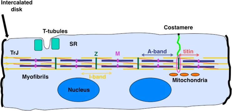
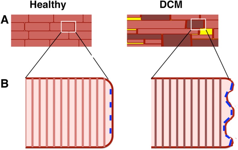
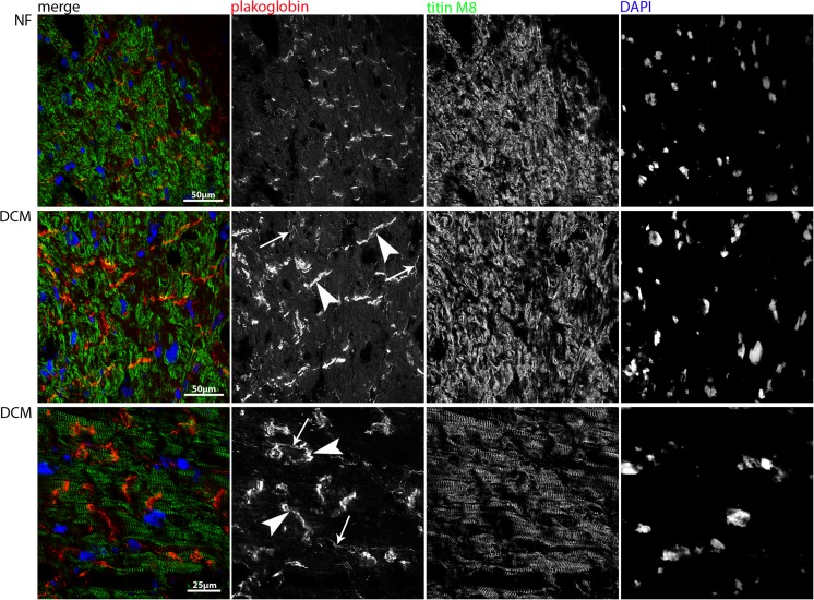
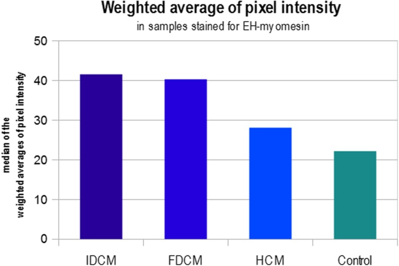
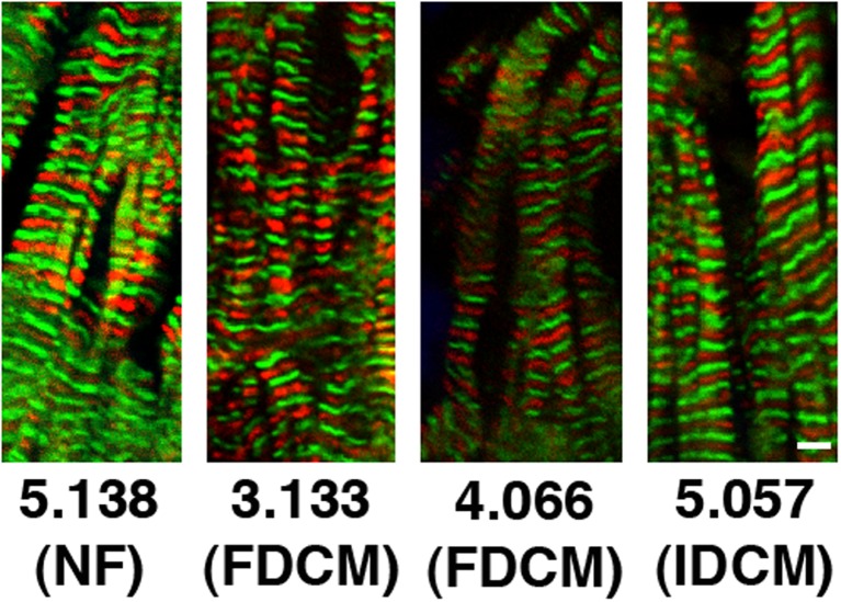
Hypertrophic cardiomyopathy is characterised by a histological phenotype of myocyte disarray, but heart tissue samples from patients with dilated cardiomyopathy (DCM) often look comparatively similar to those from healthy individuals apart from conspicuous regions of fibrosis and necrosis. We have previously investigated subcellular alterations in the cytoarchitecture of mouse models of dilated cardiomyopathy and found that both the organisation and composition of the intercalated disc, i.e. the specialised type of cell-cell contact in the heart, is altered. There is also is a change in the composition of the M-band of the sarcomere due to an expression shift towards the more extensible embryonic heart (EH)-myomesin isoform. Analysis of human samples from the Sydney Human Heart Tissue Bank have revealed similar structural findings and also provided evidence for a dramatic change in overall cardiomyocyte size control, which has also been seen in the mouse. Together these changes in cytoarchitecture probably contribute to the decreased functional output that is seen in DCM.

摘要

肥厚型心肌病的组织学表型特征为心肌细胞排列紊乱,但扩张型心肌病(DCM)患者的心脏组织样本,除了有明显的纤维化和坏死区域外,通常看起来与健康个体的样本较为相似。我们之前研究了扩张型心肌病小鼠模型细胞结构的亚细胞改变,发现闰盘(即心脏中特殊类型的细胞间连接)的组织结构和组成均发生了改变。由于向更具伸展性的胚胎心脏(EH)-肌间蛋白异构体的表达转变,肌节M带的组成也发生了变化。对悉尼人类心脏组织库的人类样本分析揭示了类似的结构发现,同时也为心肌细胞整体大小控制的显著变化提供了证据,这种变化在小鼠中也有观察到。这些细胞结构的变化共同作用,可能导致了扩张型心肌病中所见的功能输出下降。

https://cdn.ncbi.nlm.nih.gov/pmc/blobs/2213/5430298/e2665ee67a63/12551_2014_146_Fig7_HTML.jpg
https://cdn.ncbi.nlm.nih.gov/pmc/blobs/2213/5430298/3dbca10c842f/12551_2014_146_Fig1_HTML.jpg
https://cdn.ncbi.nlm.nih.gov/pmc/blobs/2213/5430298/1d5aa0945ab6/12551_2014_146_Fig2_HTML.jpg
https://cdn.ncbi.nlm.nih.gov/pmc/blobs/2213/5430298/9d053ee4c2d9/12551_2014_146_Fig3_HTML.jpg
https://cdn.ncbi.nlm.nih.gov/pmc/blobs/2213/5430298/2f40c25c2ab4/12551_2014_146_Fig4_HTML.jpg
https://cdn.ncbi.nlm.nih.gov/pmc/blobs/2213/5430298/69dd54cfe559/12551_2014_146_Fig5_HTML.jpg
https://cdn.ncbi.nlm.nih.gov/pmc/blobs/2213/5430298/a184d4c4b41b/12551_2014_146_Fig6_HTML.jpg
https://cdn.ncbi.nlm.nih.gov/pmc/blobs/2213/5430298/e2665ee67a63/12551_2014_146_Fig7_HTML.jpg

相似文献

1
Adaptations of cytoarchitecture in human dilated cardiomyopathy.人类扩张型心肌病的细胞结构适应性改变。
Biophys Rev. 2015 Mar;7(1):25-32. doi: 10.1007/s12551-014-0146-2. Epub 2014 Dec 11.
2
EH-myomesin splice isoform is a novel marker for dilated cardiomyopathy.EH-myomesin 拼接异构体是扩张型心肌病的一个新标志物。
Basic Res Cardiol. 2011 Mar;106(2):233-47. doi: 10.1007/s00395-010-0131-2. Epub 2010 Nov 11.
3
Disturbed Desmoglein-2 in the intercalated disc of pediatric patients with dilated cardiomyopathy.扩张型心肌病患儿闰盘中桥粒芯糖蛋白-2紊乱
Hum Pathol. 2017 Sep;67:101-108. doi: 10.1016/j.humpath.2017.07.012. Epub 2017 Jul 29.
4
Myofilament Remodeling and Function Is More Impaired in Peripartum Cardiomyopathy Compared with Dilated Cardiomyopathy and Ischemic Heart Disease.与扩张型心肌病和缺血性心脏病相比,围产期心肌病的肌丝重塑和功能受损更严重。
Am J Pathol. 2017 Dec;187(12):2645-2658. doi: 10.1016/j.ajpath.2017.08.022. Epub 2017 Sep 19.
5
Cardiac cytoarchitecture - why the "hardware" is important for heart function!心脏细胞结构——为何“硬件”对心脏功能至关重要!
Biochim Biophys Acta. 2016 Jul;1863(7 Pt B):1857-63. doi: 10.1016/j.bbamcr.2015.11.006. Epub 2015 Nov 11.
6
Ablation of palladin in adult heart causes dilated cardiomyopathy associated with intercalated disc abnormalities.成年心脏中 paladin 的消融导致扩张型心肌病,伴有闰盘异常。
Elife. 2023 Mar 16;12:e78629. doi: 10.7554/eLife.78629.
7
Oxidative Stress in Dilated Cardiomyopathy Caused by MYBPC3 Mutation.由MYBPC3突变引起的扩张型心肌病中的氧化应激
Oxid Med Cell Longev. 2015;2015:424751. doi: 10.1155/2015/424751. Epub 2015 Oct 5.
8
Affimers targeting proteins in the cardiomyocyte Z-disc: Novel tools that improve imaging of heart tissue.靶向心肌细胞Z盘蛋白的确认剂:改善心脏组织成像的新型工具。
Front Cardiovasc Med. 2023 Feb 14;10:1094563. doi: 10.3389/fcvm.2023.1094563. eCollection 2023.
9
Familial dilated cardiomyopathy caused by an alpha-tropomyosin mutation: the distinctive natural history of sarcomeric dilated cardiomyopathy.由α-原肌球蛋白突变引起的家族性扩张型心肌病:肌节性扩张型心肌病的独特自然病史。
J Am Coll Cardiol. 2010 Jan 26;55(4):320-9. doi: 10.1016/j.jacc.2009.11.017.
10
A mutation in the glutamate-rich region of RNA-binding motif protein 20 causes dilated cardiomyopathy through missplicing of titin and impaired Frank-Starling mechanism.RNA 结合蛋白 20 的谷氨酸丰富区的突变通过肌联蛋白的错剪接和弗兰克-斯塔尔机制受损导致扩张型心肌病。
Cardiovasc Res. 2016 Oct;112(1):452-63. doi: 10.1093/cvr/cvw192. Epub 2016 Aug 5.

引用本文的文献

1
Importance of N2BA Titin in Maintaining Cardiac Homeostasis and Its Role in Dilated Cardiomyopathy.N2BA 肌联蛋白在维持心脏稳态中的重要性及其在扩张型心肌病中的作用
Circ Heart Fail. 2025 Mar;18(3):e012083. doi: 10.1161/CIRCHEARTFAILURE.124.012083. Epub 2025 Feb 11.
2
Critical contribution of mitochondria in the development of cardiomyopathy linked to desmin mutation.线粒体在连接桥粒芯糖蛋白基因突变型心肌病发病机制中的关键作用
Stem Cell Res Ther. 2024 Jan 2;15(1):10. doi: 10.1186/s13287-023-03619-7.
3
Affimers targeting proteins in the cardiomyocyte Z-disc: Novel tools that improve imaging of heart tissue.

本文引用的文献

1
The formin FHOD1 in cardiomyocytes.心肌细胞中的formin FHOD1蛋白。
Anat Rec (Hoboken). 2014 Sep;297(9):1560-70. doi: 10.1002/ar.22984.
2
N-cadherin/catenin complex as a master regulator of intercalated disc function.N-钙黏蛋白/连环蛋白复合物作为闰盘功能的主要调节因子。
Cell Commun Adhes. 2014 Jun;21(3):169-79. doi: 10.3109/15419061.2014.908853. Epub 2014 Apr 28.
3
Cardiomyocyte growth and sarcomerogenesis at the intercalated disc.闰盘处的心肌细胞生长和肌节生成。
靶向心肌细胞Z盘蛋白的确认剂:改善心脏组织成像的新型工具。
Front Cardiovasc Med. 2023 Feb 14;10:1094563. doi: 10.3389/fcvm.2023.1094563. eCollection 2023.
4
"Meet the Editor Series"-Elisabeth Ehler.“编辑见面会系列”——伊丽莎白·埃勒
Biophys Rev. 2021 Sep 3;13(5):579-581. doi: 10.1007/s12551-021-00830-9. eCollection 2021 Oct.
5
Nanoscale regulation of L-type calcium channels differentiates between ischemic and dilated cardiomyopathies.纳米级调控 L 型钙通道可区分缺血性和扩张型心肌病。
EBioMedicine. 2020 Jul;57:102845. doi: 10.1016/j.ebiom.2020.102845. Epub 2020 Jun 21.
6
A note of appreciation for Prof. Cristobal dos Remedios on behalf of Biophysical Reviews.代表《生物物理评论》向克里斯托瓦尔·多斯·雷梅迪奥斯教授致以感谢之意。
Biophys Rev. 2019 Apr;11(2):129-130. doi: 10.1007/s12551-019-00505-6. Epub 2019 Feb 19.
7
Non-sarcomeric causes of heart failure: a Sydney Heart Bank perspective.心力衰竭的非肌节性病因:悉尼心脏库观点
Biophys Rev. 2018 Aug;10(4):949-954. doi: 10.1007/s12551-018-0441-4. Epub 2018 Jul 18.
8
Non-sarcomeric causes of heart failure.心力衰竭的非肌节性病因。
Biophys Rev. 2018 Aug;10(4):943-947. doi: 10.1007/s12551-018-0444-1. Epub 2018 Jul 14.
9
Label-free Imaging of Myocardial Remodeling in Atrial Fibrillation Using Nonlinear Optical Microscopy: A Feasibility Study.使用非线性光学显微镜对心房颤动中心肌重塑进行无标记成像:一项可行性研究。
J Atr Fibrillation. 2018 Feb 28;10(5):1644. doi: 10.4022/jafib.1644. eCollection 2018 Feb.
10
Actin-associated proteins and cardiomyopathy-the 'unknown' beyond troponin and tropomyosin.肌动蛋白相关蛋白与心肌病——肌钙蛋白和原肌球蛋白之外的“未知因素”
Biophys Rev. 2018 Aug;10(4):1121-1128. doi: 10.1007/s12551-018-0428-1. Epub 2018 Jun 5.
Cell Mol Life Sci. 2014 Jan;71(1):165-81. doi: 10.1007/s00018-013-1374-5. Epub 2013 May 26.
4
Truncations of titin causing dilated cardiomyopathy.导致扩张型心肌病的肌联蛋白截短。
N Engl J Med. 2012 Feb 16;366(7):619-28. doi: 10.1056/NEJMoa1110186.
5
Unequal allelic expression of wild-type and mutated β-myosin in familial hypertrophic cardiomyopathy.家族性肥厚型心肌病中野生型和突变型β-肌球蛋白的等位基因表达不等。
Basic Res Cardiol. 2011 Nov;106(6):1041-55. doi: 10.1007/s00395-011-0205-9. Epub 2011 Jul 19.
6
Cardiac-specific NRAP overexpression causes right ventricular dysfunction in mice.心肌特异性 NRAP 过表达导致小鼠右心室功能障碍。
Exp Cell Res. 2011 May 1;317(8):1226-37. doi: 10.1016/j.yexcr.2011.01.020. Epub 2011 Jan 26.
7
Formin follows function: a muscle-specific isoform of FHOD3 is regulated by CK2 phosphorylation and promotes myofibril maintenance.形式追随功能:FKBP12.6 同种型 FHOD3 通过 CK2 磷酸化调节,促进肌原纤维的维持。
J Cell Biol. 2010 Dec 13;191(6):1159-72. doi: 10.1083/jcb.201005060.
8
EH-myomesin splice isoform is a novel marker for dilated cardiomyopathy.EH-myomesin 拼接异构体是扩张型心肌病的一个新标志物。
Basic Res Cardiol. 2011 Mar;106(2):233-47. doi: 10.1007/s00395-010-0131-2. Epub 2010 Nov 11.
9
Modulation of human embryonic stem cell-derived cardiomyocyte growth: a testbed for studying human cardiac hypertrophy?人胚胎干细胞源性心肌细胞生长的调控:研究人类心肌肥厚的试验平台?
J Mol Cell Cardiol. 2011 Feb;50(2):367-76. doi: 10.1016/j.yjmcc.2010.10.029. Epub 2010 Nov 1.
10
Investigation of changes in skeletal muscle alpha-actin expression in normal and pathological human and mouse hearts.研究正常人和病理人心肌中骨骼肌α-肌动蛋白表达的变化。
J Muscle Res Cell Motil. 2010 Sep;31(3):207-14. doi: 10.1007/s10974-010-9224-7. Epub 2010 Aug 13.