Department of Cardiovascular Sciences, Imperial Centre for Translational and Experimental Medicine, National Heart and Lung Institute, Imperial College London, London W120NN, UK.
Department of Biomedical Engineering and Alliance for Cardiovascular Diagnostic and Treatment Innovation, Johns Hopkins University, Baltimore, MD 21218, USA.
EBioMedicine. 2020 Jul;57:102845. doi: 10.1016/j.ebiom.2020.102845. Epub 2020 Jun 21.
Subcellular localization and function of L-type calcium channels (LTCCs) play an important role in regulating contraction of cardiomyocytes. Understanding how this is affected by the disruption of transverse tubules during heart failure could lead to new insights into the disease.
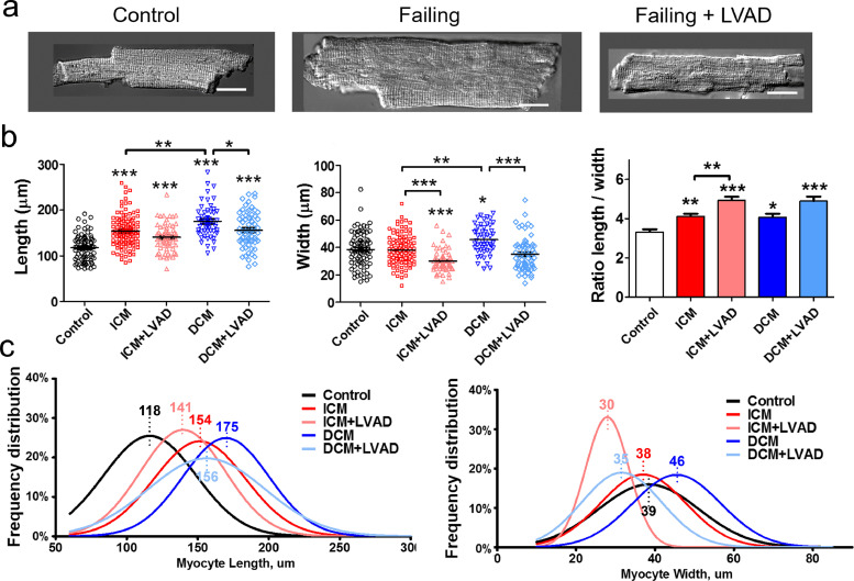
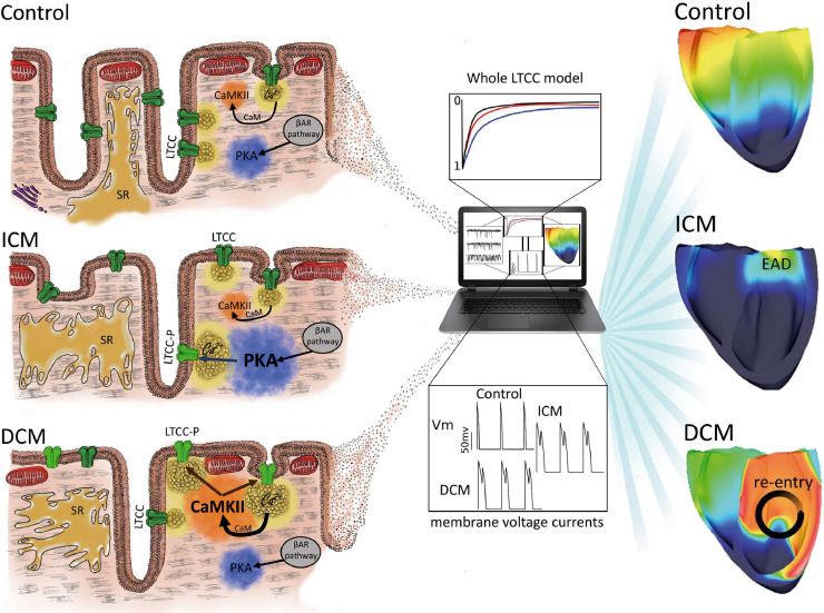
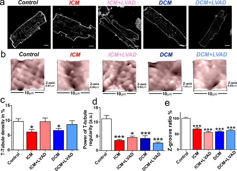
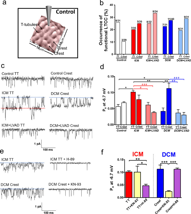
Cardiomyocytes were isolated from healthy donor hearts, as well as from patients with cardiomyopathies and with left ventricular assist devices. Scanning ion conductance and confocal microscopy was used to study membrane structures in the cells. Super-resolution scanning patch-clamp was used to examine LTCC function in different microdomains. Computational modeling predicted the impact of these changes to arrhythmogenesis at the whole-heart level.
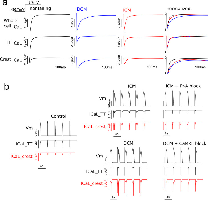
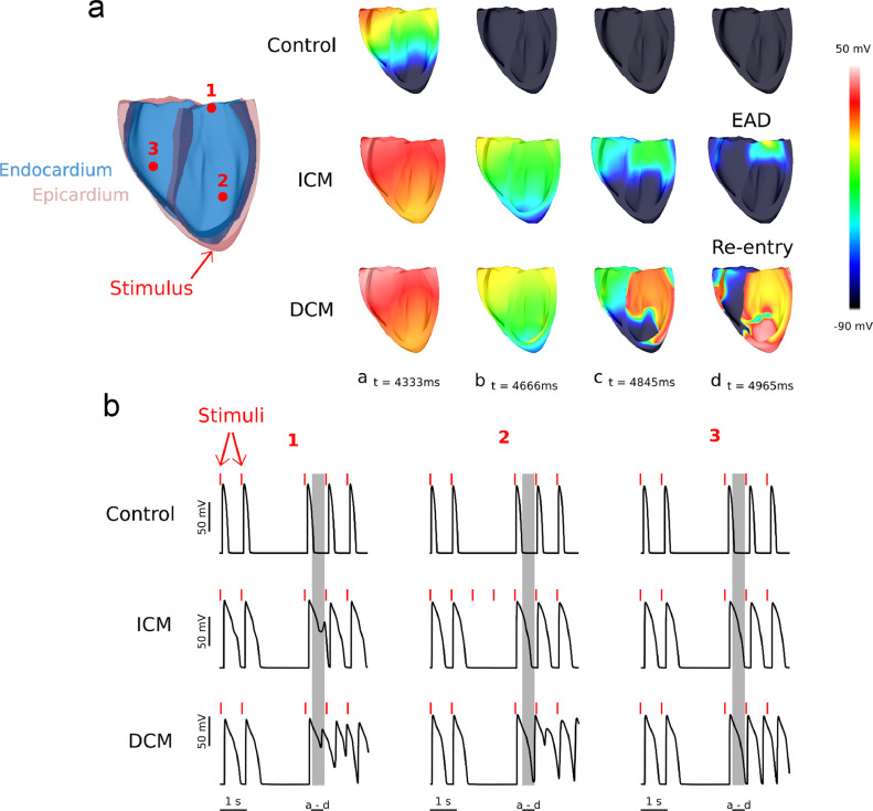
We showed that loss of structural organization in failing myocytes leads to re-distribution of functional LTCCs from the T-tubules to the sarcolemma. In ischemic cardiomyopathy, the increased LTCC open probability in the T-tubules depends on the phosphorylation by protein kinase A, whereas in dilated cardiomyopathy, the increased LTCC opening probability in the sarcolemma results from enhanced phosphorylation by calcium-calmodulin kinase II. LVAD implantation corrected LTCCs pathophysiological activity, although it did not improve their distribution. Using computational modeling in a 3D anatomically-realistic human ventricular model, we showed how LTCC location and activity can trigger heart rhythm disorders of different severity.
Our findings demonstrate that LTCC redistribution and function differentiate between disease aetiologies. The subcellular changes observed in specific microdomains could be the consequence of the action of distinct protein kinases.
This work was supported by NIH grant (ROI-HL 126802 to NT-JG) and British Heart Foundation (grant RG/17/13/33173 to JG, project grant PG/16/17/32069 to RAC). Funders had no role in study design, data collection, data analysis, interpretation, writing of the report.
L 型钙通道(LTCC)的亚细胞定位和功能在调节心肌细胞收缩中起着重要作用。了解在心力衰竭过程中横管破坏如何影响 LTCC,可能会为该疾病提供新的见解。
从健康供体心脏以及心肌病和左心室辅助设备患者中分离心肌细胞。扫描离子电导和共焦显微镜用于研究细胞中的膜结构。超分辨率扫描贴附式膜片钳用于研究不同微域中 LTCC 的功能。计算模型预测了这些变化对整个心脏水平心律失常发生的影响。
我们表明,衰竭心肌细胞中结构组织的丧失导致功能性 LTCC 从横管重新分布到肌小节。在缺血性心肌病中,T 管中 LTCC 开放概率的增加取决于蛋白激酶 A 的磷酸化,而在扩张型心肌病中,肌小节中 LTCC 开放概率的增加则源于钙调蛋白激酶 II 的增强磷酸化。LVAD 植入虽然纠正了 LTCC 的病理生理学活性,但并未改善其分布。使用计算模型在 3D 解剖逼真的人类心室模型中,我们展示了 LTCC 的位置和活性如何引发不同严重程度的心律紊乱。
我们的研究结果表明,LTCC 的重新分布和功能区分了不同的疾病病因。在特定微域中观察到的亚细胞变化可能是不同蛋白激酶作用的结果。
这项工作得到了 NIH 拨款(ROI-HL 126802 给 NT-JG)和英国心脏基金会(PG/16/17/32069 给 JG,项目拨款 RG/17/13/33173 给 RAC)的支持。资助者在研究设计、数据收集、数据分析、解释、报告撰写方面没有作用。