Chung Shinjae, Weber Franz, Zhong Peng, Tan Chan Lek, Nguyen Thuc Nghi, Beier Kevin T, Hörmann Nikolai, Chang Wei-Cheng, Zhang Zhe, Do Johnny Phong, Yao Shenqin, Krashes Michael J, Tasic Bosiljka, Cetin Ali, Zeng Hongkui, Knight Zachary A, Luo Liqun, Dan Yang
Division of Neurobiology, Department of Molecular and Cell Biology, Helen Wills Neuroscience Institute, Howard Hughes Medical Institute, University of California, Berkeley, California 94720, USA.
Department of Physiology, University of California, San Francisco, San Francisco, California 94158, USA.
Nature. 2017 May 25;545(7655):477-481. doi: 10.1038/nature22350. Epub 2017 May 17.
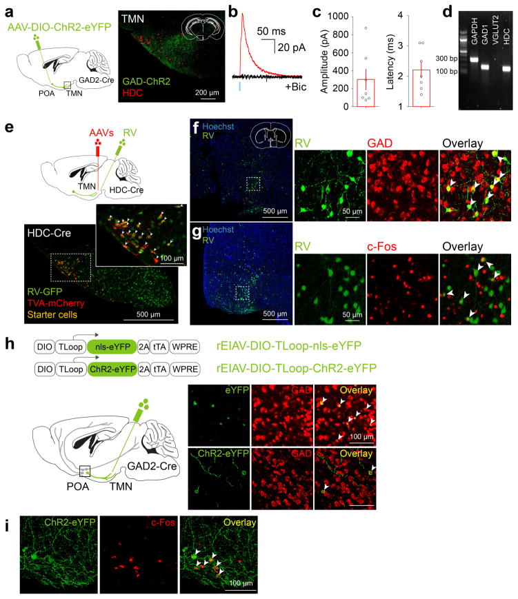
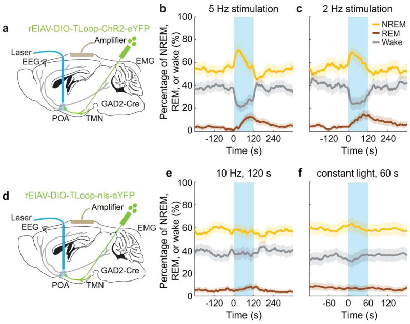
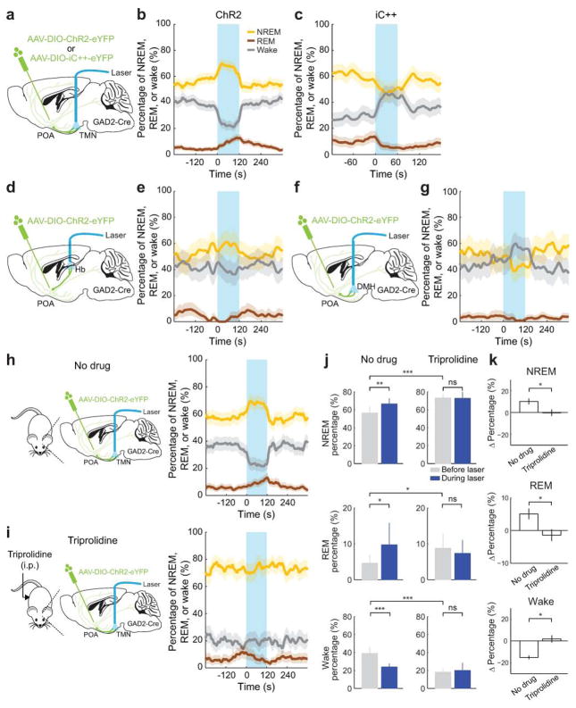
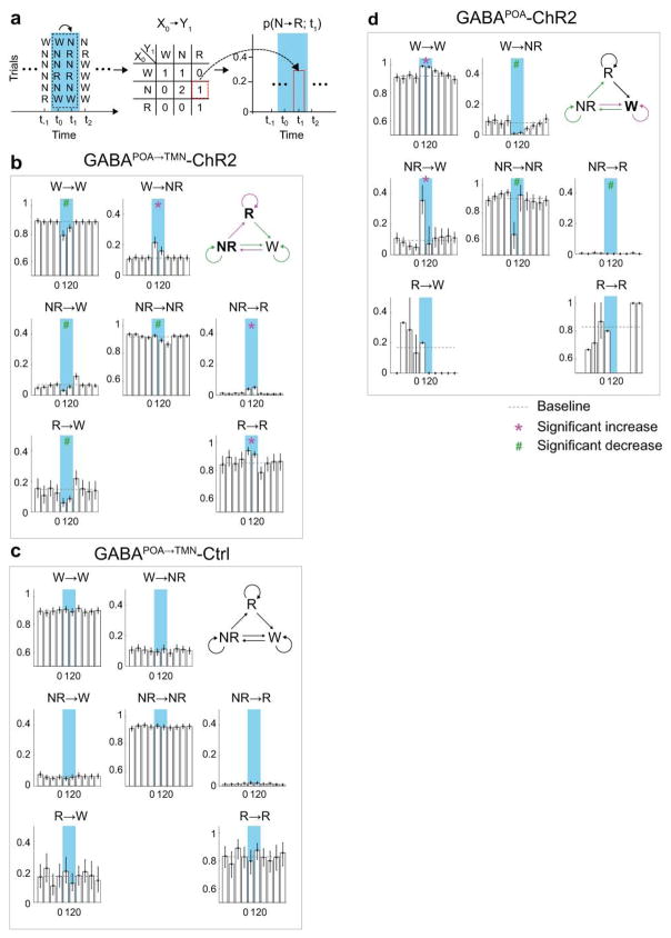
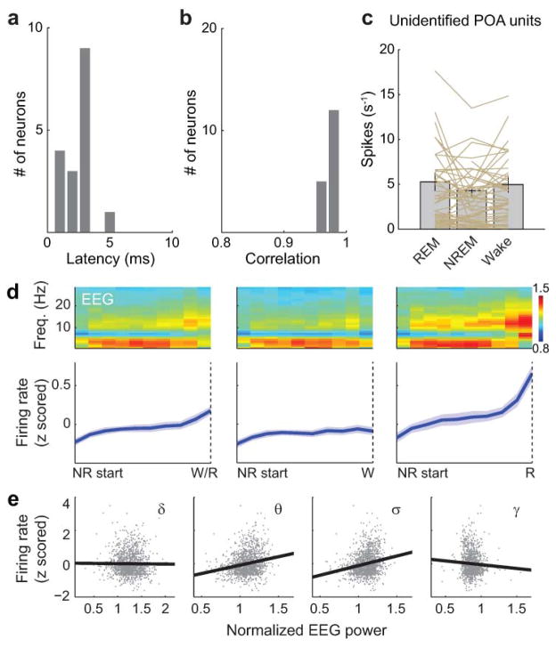
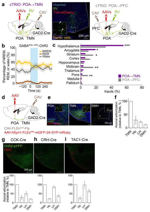
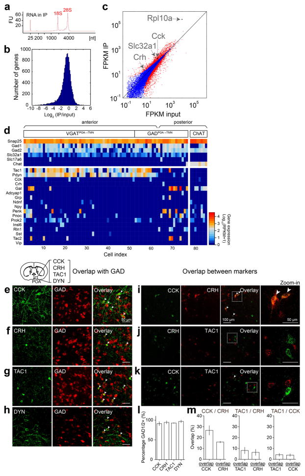
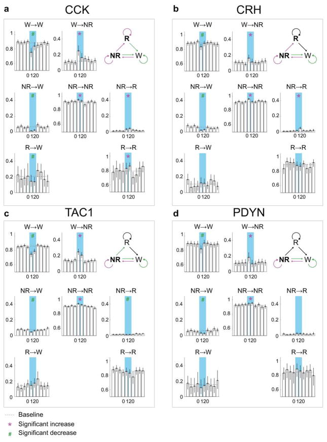
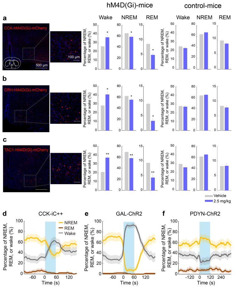
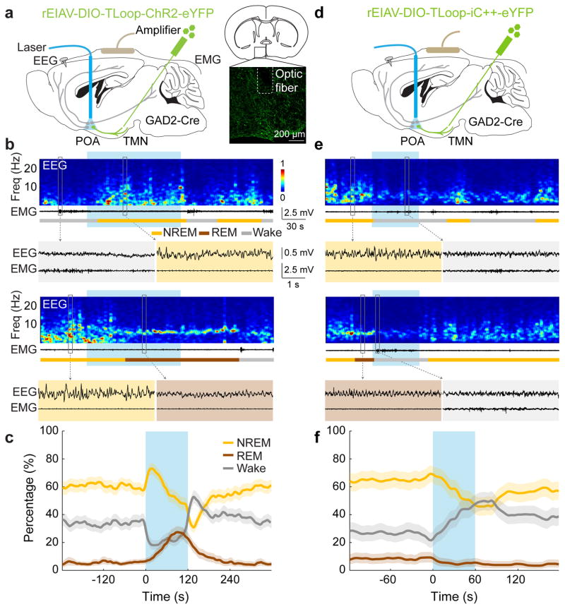
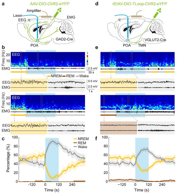
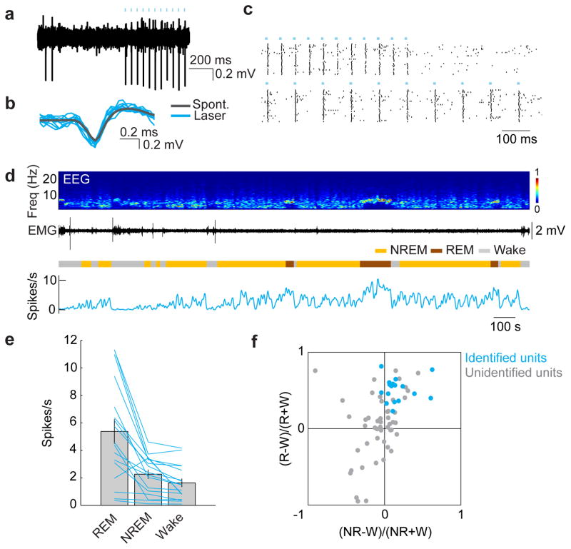
In humans and other mammalian species, lesions in the preoptic area of the hypothalamus cause profound sleep impairment, indicating a crucial role of the preoptic area in sleep generation. However, the underlying circuit mechanism remains poorly understood. Electrophysiological recordings and c-Fos immunohistochemistry have shown the existence of sleep-active neurons in the preoptic area, especially in the ventrolateral preoptic area and median preoptic nucleus. Pharmacogenetic activation of c-Fos-labelled sleep-active neurons has been shown to induce sleep. However, the sleep-active neurons are spatially intermingled with wake-active neurons, making it difficult to target the sleep neurons specifically for circuit analysis. Here we identify a population of preoptic area sleep neurons on the basis of their projection target and discover their molecular markers. Using a lentivirus expressing channelrhodopsin-2 or a light-activated chloride channel for retrograde labelling, bidirectional optogenetic manipulation, and optrode recording, we show that the preoptic area GABAergic neurons projecting to the tuberomammillary nucleus are both sleep active and sleep promoting. Furthermore, translating ribosome affinity purification and single-cell RNA sequencing identify candidate markers for these neurons, and optogenetic and pharmacogenetic manipulations demonstrate that several peptide markers (cholecystokinin, corticotropin-releasing hormone, and tachykinin 1) label sleep-promoting neurons. Together, these findings provide easy genetic access to sleep-promoting preoptic area neurons and a valuable entry point for dissecting the sleep control circuit.
在人类和其他哺乳动物中,下丘脑视前区的损伤会导致严重的睡眠障碍,这表明视前区在睡眠产生中起着关键作用。然而,其潜在的神经回路机制仍知之甚少。电生理记录和c-Fos免疫组织化学显示,视前区存在睡眠激活神经元,特别是在腹外侧视前区和视前正中核。c-Fos标记的睡眠激活神经元的光遗传学激活已被证明可诱导睡眠。然而,睡眠激活神经元在空间上与觉醒激活神经元相互交织,这使得难以特异性地靶向睡眠神经元进行神经回路分析。在这里,我们根据视前区睡眠神经元的投射靶点鉴定出一群此类神经元,并发现了它们的分子标记。通过使用表达通道视紫红质-2的慢病毒或光激活氯离子通道进行逆行标记、双向光遗传学操作和光电极记录,我们发现投射到结节乳头体核的视前区GABA能神经元既具有睡眠激活作用,又能促进睡眠。此外,翻译核糖体亲和纯化和单细胞RNA测序确定了这些神经元的候选标记,光遗传学和药理学操作表明,几种肽类标记物(胆囊收缩素、促肾上腺皮质激素释放激素和速激肽1)标记了促进睡眠的神经元。总之,这些发现为促进睡眠的视前区神经元提供了便捷的基因研究途径,并为剖析睡眠控制神经回路提供了一个有价值的切入点。