Ciria María, García Nahuel A, Ontoria-Oviedo Imelda, González-King Hernán, Carrero Rubén, De La Pompa José Luis, Montero José Anastasio, Sepúlveda Pilar
1 Regenerative Medicine and Heart Transplantation Unit, Instituto de Investigación Sanitaria La Fe , Valencia, Spain .
2 Joint Unit for Cardiovascular Repair, Instituto de Investigación Sanitaria La Fe-Centro de Investigación Príncipe Felipe , Valencia, Spain .
Stem Cells Dev. 2017 Jul 1;26(13):973-985. doi: 10.1089/scd.2016.0331. Epub 2017 May 18.
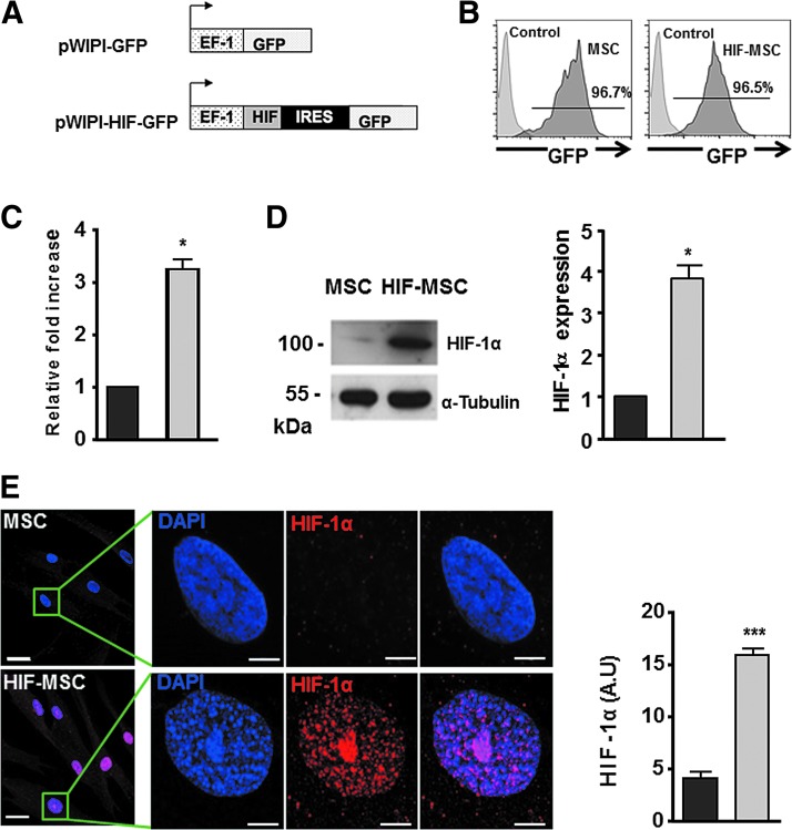
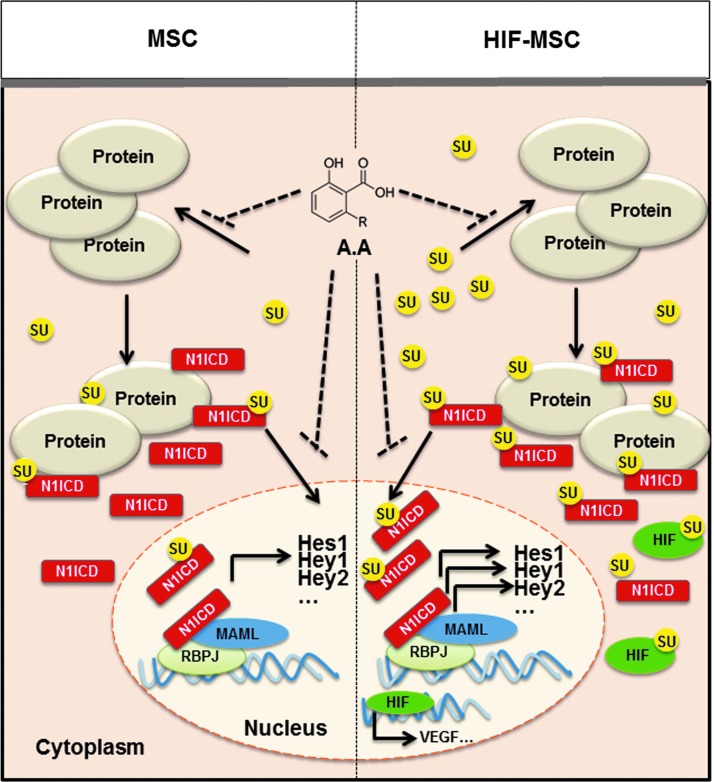
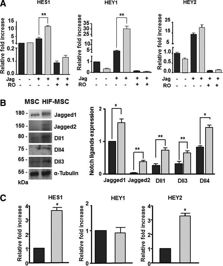
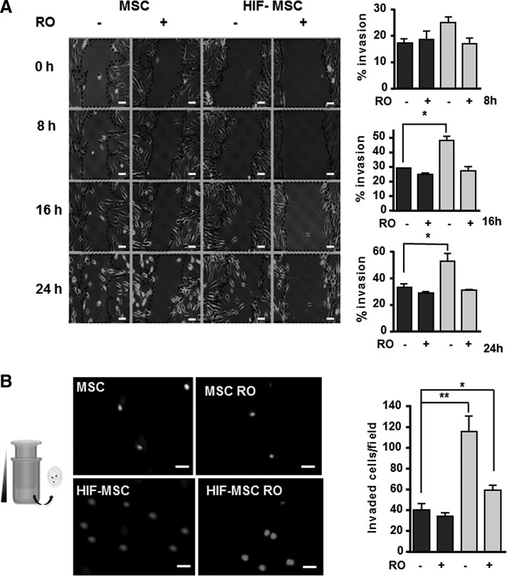
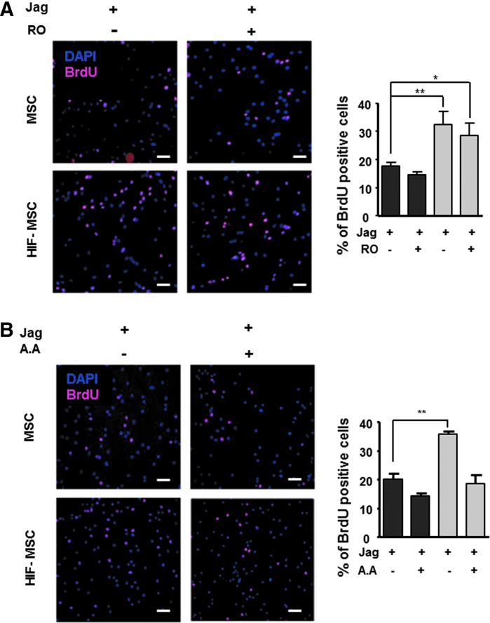
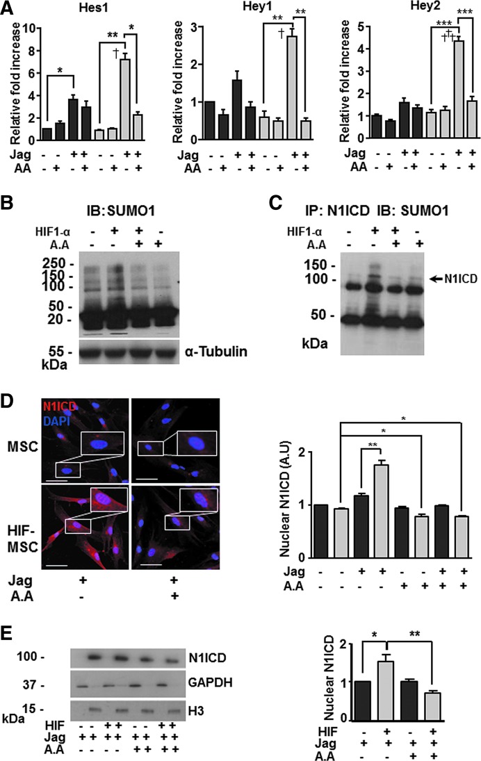
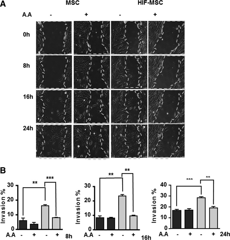
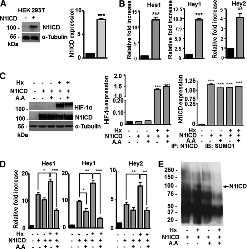
Mesenchymal stem cells (MSCs) are effective in treating several pathologies. We and others have demonstrated that hypoxia or hypoxia-inducible factor 1 alpha (HIF-1α) stabilization improves several MSC functions, including cell adhesion, migration, and proliferation, thereby increasing their therapeutic potential. To further explore the mechanisms induced by HIF-1α in MSCs, we studied its relationship with Notch signaling and observed that overexpression of HIF-1α in MSCs increased protein levels of the Notch ligands Jagged 1-2 and Delta-like (Dll)1, Dll3, and Dll4 and potentiated Notch signaling only when this pathway was activated. Crosstalk between HIF and Notch resulted in Notch-dependent migration and spreading of MSCs, which was abolished by γ-secretase inhibition. However, the HIF-1-induced increase in MSC proliferation was independent of Notch signaling. The ubiquitin family member, small ubiquitin-like modifier (SUMO), has important functions in many cellular processes and increased SUMO1 protein levels have been reported in hypoxia. To investigate the potential involvement of SUMOylation in HIF/Notch crosstalk, we measured general SUMOylation levels and observed increased SUMOylation in HIF-1-expressing MSCs. Moreover, proliferation and migration of MSCs were reduced in the presence of a SUMOylation inhibitor, and this effect was particularly robust in HIF-MSCs. Immunoprecipitation studies demonstrated SUMOylation of the intracellular domain of Notch1 (N1ICD) in HIF-1-expressing MSCs, which contributed to Notch pathway activation and resulted in increased levels of N1ICD nuclear translocation as assessed by subcellular fractionation. SUMOylation of N1ICD was also observed in HEK293T cells with stabilized HIF-1α expression, suggesting that this is a common mechanism in eukaryotic cells. In summary, we describe, for the first time, SUMOylation of N1ICD, which is potentiated by HIF signaling. These phenomena could be relevant for the therapeutic effects of MSCs in hypoxia or under conditions of HIF stabilization.
间充质干细胞(MSCs)在治疗多种疾病方面具有有效性。我们和其他研究人员已证明,缺氧或缺氧诱导因子1α(HIF-1α)的稳定化可改善多种MSC功能,包括细胞黏附、迁移和增殖,从而增强其治疗潜力。为了进一步探究HIF-1α在MSCs中诱导的机制,我们研究了其与Notch信号通路的关系,并观察到MSCs中HIF-1α的过表达增加了Notch配体Jagged 1-2和Delta样(Dll)1、Dll3及Dll4的蛋白水平,且仅在该信号通路被激活时增强了Notch信号。HIF与Notch之间的相互作用导致了MSCs依赖Notch的迁移和铺展,而γ-分泌酶抑制可消除这种现象。然而,HIF-1诱导的MSCs增殖增加与Notch信号通路无关。泛素家族成员小泛素样修饰物(SUMO)在许多细胞过程中具有重要功能,并且已有报道称缺氧时SUMO1蛋白水平升高。为了研究SUMO化在HIF/Notch相互作用中的潜在作用,我们检测了总体SUMO化水平,并观察到表达HIF-1的MSCs中SUMO化增加。此外,在存在SUMO化抑制剂的情况下,MSCs的增殖和迁移减少,并且这种效应在HIF-MSCs中尤为明显。免疫沉淀研究表明,在表达HIF-1的MSCs中,Notch1的细胞内结构域(N1ICD)发生了SUMO化,这有助于Notch信号通路的激活,并通过亚细胞分级分离评估发现导致N1ICD核转位水平增加。在稳定表达HIF-1α的HEK293T细胞中也观察到了N1ICD的SUMO化,这表明这是真核细胞中的一种常见机制。总之,我们首次描述了N1ICD的SUMO化,它由HIF信号增强。这些现象可能与MSCs在缺氧或HIF稳定化条件下的治疗效果相关。