• 文献检索
  • 文档翻译
  • 深度研究
  • 学术资讯
  • Suppr Zotero 插件Zotero 插件
  • 邀请有礼
  • 套餐&价格
  • 历史记录
应用&插件
Suppr Zotero 插件Zotero 插件浏览器插件Mac 客户端Windows 客户端微信小程序
定价
高级版会员购买积分包购买API积分包
服务
文献检索文档翻译深度研究API 文档MCP 服务
关于我们
关于 Suppr公司介绍联系我们用户协议隐私条款
关注我们

Suppr 超能文献

核心技术专利:CN118964589B侵权必究
粤ICP备2023148730 号-1Suppr @ 2026

文献检索

告别复杂PubMed语法,用中文像聊天一样搜索,搜遍4000万医学文献。AI智能推荐,让科研检索更轻松。

立即免费搜索

文件翻译

保留排版,准确专业,支持PDF/Word/PPT等文件格式,支持 12+语言互译。

免费翻译文档

深度研究

AI帮你快速写综述,25分钟生成高质量综述,智能提取关键信息,辅助科研写作。

立即免费体验

Notch 信号通路在心肌梗死后心脏功能恢复中的作用。

The Role of the Notch Signaling Pathway in Recovery of Cardiac Function after Myocardial Infarction.

机构信息

Laboratory of Regenerative Biomedicine, Institute of Cytology, Russian Academy of Sciences, 119991 Saint-Petersburg, Russia.

出版信息

Int J Mol Sci. 2022 Oct 19;23(20):12509. doi: 10.3390/ijms232012509.

DOI:10.3390/ijms232012509
PMID:36293363
原文链接:https://pmc.ncbi.nlm.nih.gov/articles/PMC9604421/
Abstract

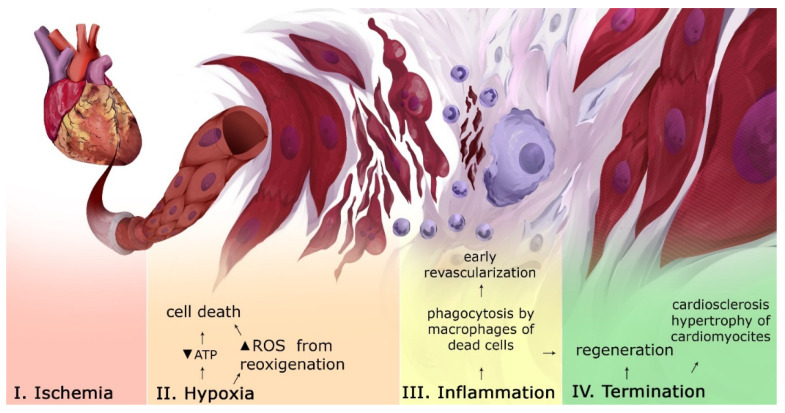
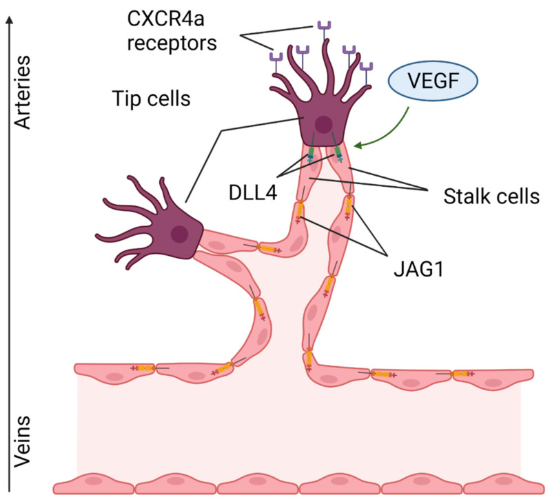
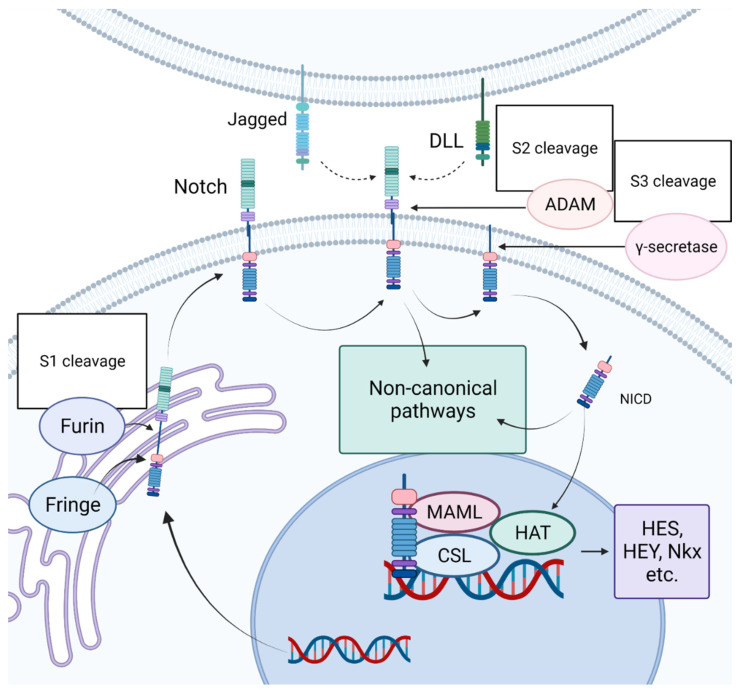
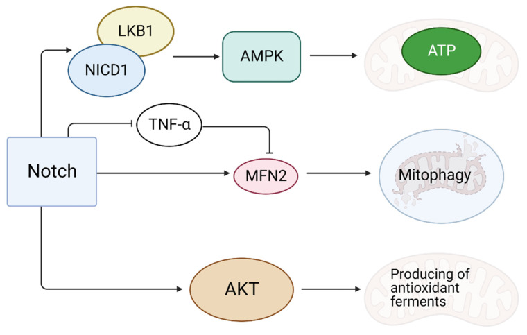
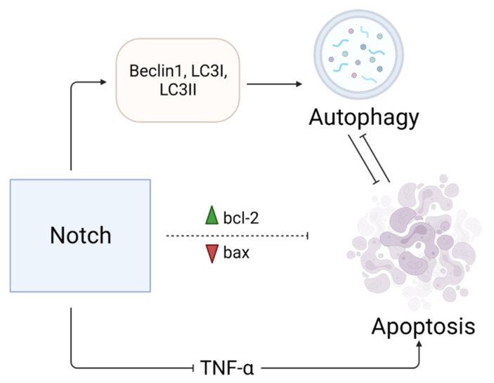
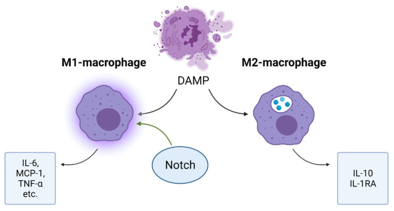
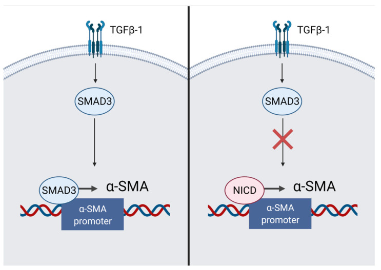
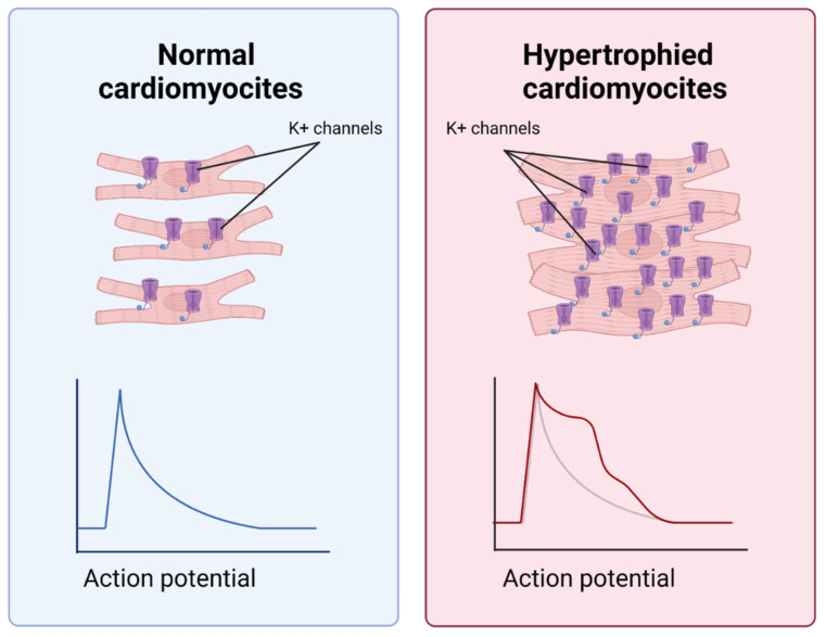
Myocardial infarction (MI) is a pathological process, evidencing as massive death of cardiomyocytes associated with hypoxic and oxidative stress. The formation of areas of fibrosis ultimately leads to heart failure. There are some mechanisms that contribute to the functional repair of the heart. In most mammals, including humans, the Notch signaling pathway has cardioprotective effects. It is involved in the formation of the heart in embryogenesis and in the restoration of cardiac function after MI due to: (1) reducing oxidative stress; (2) prevention of apoptosis; (3) regulation of inflammation; (4) containment of fibrosis and hypertrophy of cardiomyocytes; (5) tissue revascularization; and (6) regulation of proliferation and differentiation of cardiomyocytes. In addition, the Notch signaling pathway interacts with other signaling cascades involved in the pathogenesis of MI and subsequent cardiac repair. In this review, we consider the Notch signaling pathway as a potential target for therapeutic approaches aimed at improving cardiac recovery after MI.

摘要

心肌梗死(MI)是一种病理过程,表现为与缺氧和氧化应激相关的大量心肌细胞死亡。纤维化区域的形成最终导致心力衰竭。有一些机制有助于心脏的功能修复。在大多数哺乳动物中,包括人类,Notch 信号通路具有心脏保护作用。它参与胚胎心脏的形成以及 MI 后心脏功能的恢复,原因是:(1)减少氧化应激;(2)防止细胞凋亡;(3)调节炎症;(4)抑制心肌细胞纤维化和肥大;(5)组织再血管化;(6)调节心肌细胞的增殖和分化。此外,Notch 信号通路与 MI 发病机制和随后心脏修复过程中涉及的其他信号级联相互作用。在这篇综述中,我们将 Notch 信号通路视为一种有潜力的治疗靶点,旨在改善 MI 后心脏的恢复。

https://cdn.ncbi.nlm.nih.gov/pmc/blobs/7b92/9604421/9158dc557b16/ijms-23-12509-g008.jpg
https://cdn.ncbi.nlm.nih.gov/pmc/blobs/7b92/9604421/b120d3eda380/ijms-23-12509-g001.jpg
https://cdn.ncbi.nlm.nih.gov/pmc/blobs/7b92/9604421/f17a901c9e25/ijms-23-12509-g002.jpg
https://cdn.ncbi.nlm.nih.gov/pmc/blobs/7b92/9604421/78431f3a8876/ijms-23-12509-g003.jpg
https://cdn.ncbi.nlm.nih.gov/pmc/blobs/7b92/9604421/c95c8119f250/ijms-23-12509-g004.jpg
https://cdn.ncbi.nlm.nih.gov/pmc/blobs/7b92/9604421/6aa0ba5610d7/ijms-23-12509-g005.jpg
https://cdn.ncbi.nlm.nih.gov/pmc/blobs/7b92/9604421/a63b72d3cc8a/ijms-23-12509-g006.jpg
https://cdn.ncbi.nlm.nih.gov/pmc/blobs/7b92/9604421/f9c1320b2f41/ijms-23-12509-g007.jpg
https://cdn.ncbi.nlm.nih.gov/pmc/blobs/7b92/9604421/9158dc557b16/ijms-23-12509-g008.jpg

相似文献

1
The Role of the Notch Signaling Pathway in Recovery of Cardiac Function after Myocardial Infarction. Notch 信号通路在心肌梗死后心脏功能恢复中的作用。
Int J Mol Sci. 2022 Oct 19;23(20):12509. doi: 10.3390/ijms232012509.
2
Notch signaling as an important mediator of cardiac repair and regeneration after myocardial infarction.Notch 信号作为心肌梗死后心脏修复和再生的重要介质。
Trends Cardiovasc Med. 2010 Oct;20(7):228-31. doi: 10.1016/j.tcm.2011.11.006.
3
Blockade of RBP-J-Mediated Notch Signaling Pathway Exacerbates Cardiac Remodeling after Infarction by Increasing Apoptosis in Mice.阻断 RBP-J 介导的 Notch 信号通路通过增加小鼠梗死心肌细胞凋亡加重心脏重构。
Biomed Res Int. 2018 Jul 3;2018:5207031. doi: 10.1155/2018/5207031. eCollection 2018.
4
MyD88 mediated inflammatory signaling leads to CaMKII oxidation, cardiac hypertrophy and death after myocardial infarction.MyD88 介导的炎症信号导致心肌梗死后的 CaMKII 氧化、心脏肥大和死亡。
J Mol Cell Cardiol. 2012 May;52(5):1135-44. doi: 10.1016/j.yjmcc.2012.01.021. Epub 2012 Feb 3.
5
Therapeutic effect of a novel Wnt pathway inhibitor on cardiac regeneration after myocardial infarction.一种新型Wnt信号通路抑制剂对心肌梗死后心脏再生的治疗作用
Clin Sci (Lond). 2017 Dec 8;131(24):2919-2932. doi: 10.1042/CS20171256. Print 2017 Dec 15.
6
The Notch pathway controls fibrotic and regenerative repair in the adult heart.Notch信号通路控制成年心脏的纤维化和再生修复。
Eur Heart J. 2014 Aug 21;35(32):2174-85. doi: 10.1093/eurheartj/ehs269. Epub 2012 Nov 19.
7
Notch signal pathway - therapeutic target for regulation of reparative processes in the heart.Notch信号通路——心脏修复过程调控的治疗靶点。
Ter Arkh. 2018 Dec 30;90(12):112-121. doi: 10.26442/00403660.2018.12.000014.
8
Apelin-13 increases myocardial progenitor cells and improves repair postmyocardial infarction.Apelin-13 可增加心肌祖细胞数量,改善心肌梗死后的修复。
Am J Physiol Heart Circ Physiol. 2012 Sep 1;303(5):H605-18. doi: 10.1152/ajpheart.00366.2012. Epub 2012 Jun 29.
9
LncRNA Snhg1-driven self-reinforcing regulatory network promoted cardiac regeneration and repair after myocardial infarction.长链非编码 RNA Snhg1 驱动的自我增强调控网络促进心肌梗死后的心脏再生和修复。
Theranostics. 2021 Sep 13;11(19):9397-9414. doi: 10.7150/thno.57037. eCollection 2021.
10
Oestrogen Receptor β Activation Protects Against Myocardial Infarction via Notch1 Signalling.雌激素受体 β 激活通过 Notch1 信号通路保护心肌免受梗死。
Cardiovasc Drugs Ther. 2020 Apr;34(2):165-178. doi: 10.1007/s10557-020-06949-3.

引用本文的文献

1
CD4 T Cell Subsets and as Novel Biomarkers of Immune Dysregulation in Dilated Cardiomyopathy.CD4 T细胞亚群作为扩张型心肌病免疫失调的新型生物标志物
Int J Mol Sci. 2025 Aug 13;26(16):7806. doi: 10.3390/ijms26167806.
2
Carvedilol sensitizes paclitaxel-resistant gastric cancer AGS cells to paclitaxel: influences on apoptotic regulators, Notch, PI3K/AKT, ERK1/2 signaling pathways, and miR-34a expression.卡维地洛使耐紫杉醇的胃癌AGS细胞对紫杉醇敏感:对凋亡调节因子、Notch、PI3K/AKT、ERK1/2信号通路及miR-34a表达的影响
Med Oncol. 2025 Jul 30;42(9):392. doi: 10.1007/s12032-025-02966-0.
3
Heart failure, inflammation and exercise.

本文引用的文献

1
Mechanisms of Regenerative Potential Activation in Cardiac Mesenchymal Cells.心脏间充质细胞中再生潜能激活的机制
Biomedicines. 2022 May 31;10(6):1283. doi: 10.3390/biomedicines10061283.
2
The Significance of NOTCH Pathway in the Development of Fibrosis in Systemic Sclerosis.NOTCH信号通路在系统性硬化症纤维化发展中的意义
Ann Dermatol. 2019 Aug;31(4):365-371. doi: 10.5021/ad.2019.31.4.365. Epub 2019 Jul 1.
3
The interaction of Notch and Wnt signaling pathways in vertebrate regeneration.脊椎动物再生过程中Notch和Wnt信号通路的相互作用。
心力衰竭、炎症与运动
Int J Biol Sci. 2025 Apr 28;21(8):3324-3350. doi: 10.7150/ijbs.109917. eCollection 2025.
4
Inactivation of Notch1-TGF-β-Smads Signaling Pathway by Atorvastatin Improves Cardiac Function and Hemodynamic Performance in Acute Myocardial Infarction Rats.阿托伐他汀对Notch1-TGF-β-Smads信号通路的失活作用改善急性心肌梗死大鼠的心功能和血流动力学性能
J Vasc Res. 2025;62(3):133-144. doi: 10.1159/000542728. Epub 2025 Jan 27.
5
Competitive signaling and cellular communications in myocardial infarction response.心肌梗死反应中的竞争性信号传导与细胞通讯
Mol Biol Rep. 2025 Jan 16;52(1):129. doi: 10.1007/s11033-025-10236-5.
6
The relationship between inflammatory factors and heart failure: evidence based on bidirectional Mendelian randomization analysis.炎症因子与心力衰竭之间的关系:基于双向孟德尔随机化分析的证据
Front Cardiovasc Med. 2024 Dec 12;11:1378327. doi: 10.3389/fcvm.2024.1378327. eCollection 2024.
7
Berberine ameliorates septic cardiomyopathy through protecting mitochondria and upregulating Notch1 signaling in cardiomyocytes.小檗碱通过保护心肌细胞中的线粒体和上调Notch1信号通路来改善脓毒症性心肌病。
Front Pharmacol. 2024 Nov 6;15:1502354. doi: 10.3389/fphar.2024.1502354. eCollection 2024.
8
Molecular Interplay in Cardiac Fibrosis: Exploring the Functions of RUNX2, BMP2, and Notch.心脏纤维化中的分子相互作用:探索RUNX2、BMP2和Notch的功能
Rev Cardiovasc Med. 2024 Oct 16;25(10):368. doi: 10.31083/j.rcm2510368. eCollection 2024 Oct.
9
The Complex Interplay of TGF-β and Notch Signaling in the Pathogenesis of Fibrosis.TGF-β 和 Notch 信号通路在纤维化发病机制中的复杂相互作用。
Int J Mol Sci. 2024 Oct 8;25(19):10803. doi: 10.3390/ijms251910803.
10
Identification of hub modules and therapeutic targets associated with CD8T-cells in HF and their pan-cancer analysis.鉴定与心力衰竭中 CD8T 细胞相关的枢纽模块和治疗靶点及其泛癌分析。
Sci Rep. 2024 Aug 13;14(1):18823. doi: 10.1038/s41598-024-68504-6.
Cell Regen. 2021 Apr 1;10(1):11. doi: 10.1186/s13619-020-00072-2.
4
Network-based screen in iPSC-derived cells reveals therapeutic candidate for heart valve disease.基于网络的 iPSC 衍生细胞筛选揭示了心脏瓣膜疾病的治疗候选药物。
Science. 2021 Feb 12;371(6530). doi: 10.1126/science.abd0724. Epub 2020 Dec 10.
5
Cardiac Regeneration After Myocardial Infarction: an Approachable Goal.心肌梗死后的心脏再生:一个可行的目标。
Curr Cardiol Rep. 2020 Aug 10;22(10):122. doi: 10.1007/s11886-020-01361-7.
6
Inhibition of the Notch1 pathway induces peripartum cardiomyopathy.抑制 Notch1 通路可诱导围生期心肌病。
J Cell Mol Med. 2020 Jul;24(14):7907-7914. doi: 10.1111/jcmm.15423. Epub 2020 Jun 11.
7
HIF1α or mitophagy: which drives cardiomyocyte differentiation?低氧诱导因子1α(HIF1α)还是线粒体自噬:哪个驱动心肌细胞分化?
Cell Stress. 2020 May 11;4(5):95-98. doi: 10.15698/cst2020.05.219.
8
Notch1 protects against myocardial ischaemia-reperfusion injury via regulating mitochondrial fusion and function.Notch1 通过调节线粒体融合和功能来保护心肌免受缺血再灌注损伤。
J Cell Mol Med. 2020 Mar;24(5):3183-3191. doi: 10.1111/jcmm.14992. Epub 2020 Jan 23.
9
Dysregulation of Notch signaling in cardiac mesenchymal cells of patients with tetralogy of Fallot.四叶型主动脉干患者心脏间质细胞中 Notch 信号的失调。
Pediatr Res. 2020 Jul;88(1):38-47. doi: 10.1038/s41390-020-0760-6. Epub 2020 Jan 17.
10
Cardio-renal Exosomes in Myocardial Infarction Serum Regulate Proangiogenic Paracrine Signaling in Adipose Mesenchymal Stem Cells.心肌梗死后血清中的心脏-肾脏外泌体调节脂肪间充质干细胞中的促血管生成旁分泌信号。
Theranostics. 2020 Jan 1;10(3):1060-1073. doi: 10.7150/thno.37678. eCollection 2020.