Desai Pujan K, Tseng Hubert, Souza Glauco R
Nano3D Biosciences, Houston, TX 77030, USA.
Department of Internal Medicine, University of Texas Health Science Center at Houston, Houston, TX 77030, USA.
Int J Mol Sci. 2017 May 18;18(5):1085. doi: 10.3390/ijms18051085.
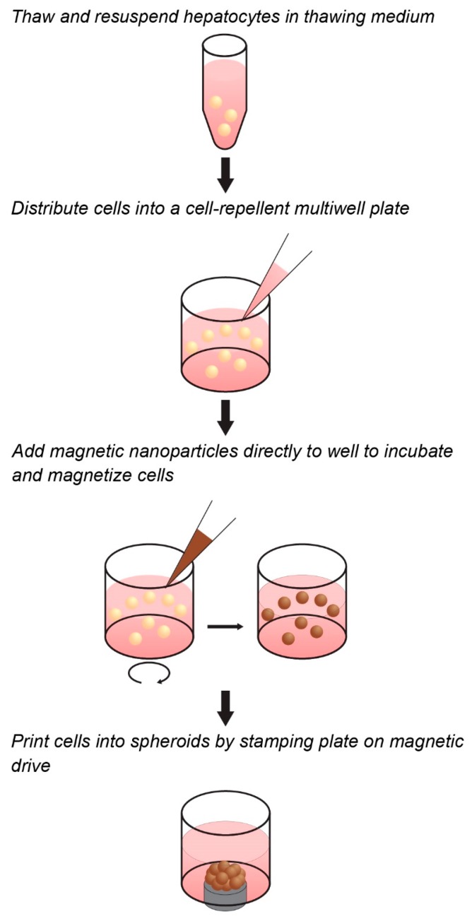
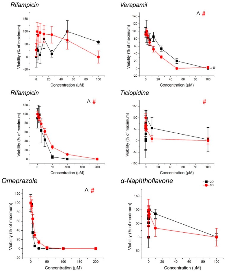
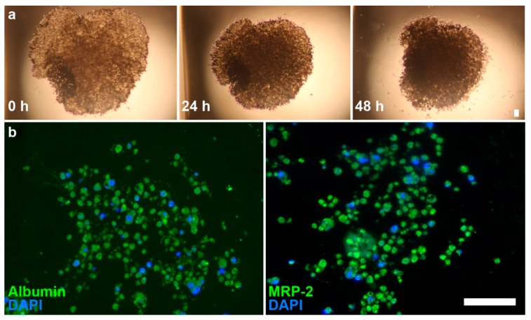
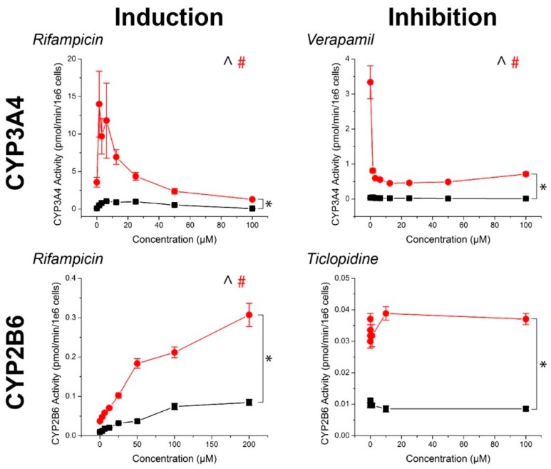
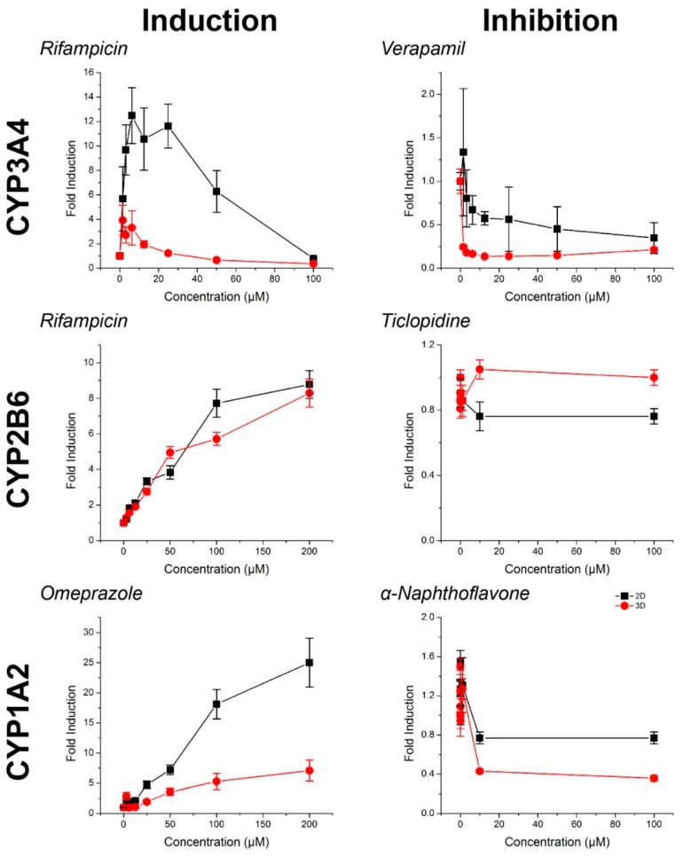
There is a significant need for in vitro methods to study drug-induced liver injury that are rapid, reproducible, and scalable for existing high-throughput systems. However, traditional monolayer and suspension cultures of hepatocytes are difficult to handle and risk the loss of phenotype. Generally, three-dimensional (3D) cell culture platforms help recapitulate native liver tissue phenotype, but suffer from technical limitations for high-throughput screening, including scalability, speed, and handling. Here, we developed a novel assay for cytochrome P450 (CYP450) induction/inhibition using magnetic 3D cell culture that overcomes the limitations of other platforms by aggregating magnetized cells with magnetic forces. With this platform, spheroids can be rapidly assembled and easily handled, while replicating native liver function. We assembled spheroids of primary human hepatocytes in a 384-well format and maintained this culture over five days, including a 72 h induction period with known CYP450 inducers/inhibitors. CYP450 activity and viability in the spheroids were assessed and compared in parallel with monolayers. CYP450 activity was induced/inhibited in spheroids as expected, separate from any toxic response. Spheroids showed a significantly higher baseline level of CYP450 activity and induction over monolayers. Positive staining in spheroids for albumin and multidrug resistance-associated protein (MRP2) indicates the preservation of hepatocyte function within spheroids. The study presents a proof-of-concept for the use of magnetic 3D cell culture for the assembly and handling of novel hepatic tissue models.
迫切需要一种体外方法来研究药物性肝损伤,该方法要快速、可重复且能适用于现有的高通量系统。然而,传统的肝细胞单层培养和悬浮培养难以操作,且存在表型丧失的风险。一般来说,三维(3D)细胞培养平台有助于重现天然肝组织表型,但在高通量筛选方面存在技术限制,包括可扩展性、速度和操作难度。在此,我们开发了一种利用磁性3D细胞培养进行细胞色素P450(CYP450)诱导/抑制的新型检测方法,该方法通过磁力聚集磁化细胞克服了其他平台的局限性。借助这个平台,球体可以快速组装且易于操作,同时能复制天然肝功能。我们以384孔板的形式组装了原代人肝细胞球体,并将这种培养维持了五天,其中包括用已知的CYP450诱导剂/抑制剂进行72小时的诱导期。对球体中的CYP450活性和活力进行了评估,并与单层培养进行了平行比较。正如预期的那样,球体中的CYP450活性被诱导/抑制,与任何毒性反应无关。球体显示出比单层培养显著更高的CYP450活性基线水平和诱导水平。球体中白蛋白和多药耐药相关蛋白(MRP2)的阳性染色表明球体中肝细胞功能得以保留。该研究为利用磁性3D细胞培养来组装和处理新型肝组织模型提供了概念验证。