Molina Rosana S, Tran Tam M, Campbell Robert E, Lambert Gerard G, Salih Anya, Shaner Nathan C, Hughes Thomas E, Drobizhev Mikhail
Department of Cell Biology & Neuroscience, Montana State University , Bozeman, Montana 59717, United States.
Department of Chemistry, University of Alberta , Edmonton, Alberta T6G 2R3, Canada.
J Phys Chem Lett. 2017 Jun 15;8(12):2548-2554. doi: 10.1021/acs.jpclett.7b00960. Epub 2017 May 25.
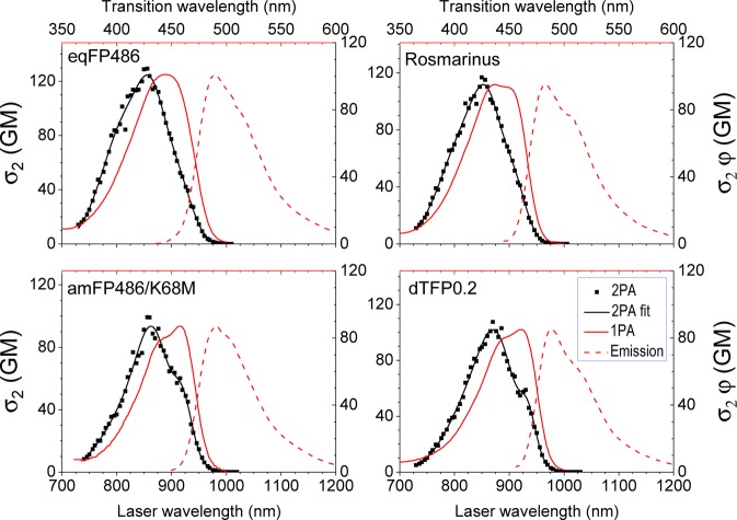
Fluorescent proteins (FPs) are indispensable markers for two-photon imaging of live tissue, especially in the brains of small model organisms. The quantity of physiologically relevant data collected, however, is limited by heat-induced damage of the tissue due to the high intensities of the excitation laser. We seek to minimize this damage by developing FPs with improved brightness. Among FPs with the same chromophore structure, the spectral properties can vary widely due to differences in the local protein environment. Using a physical model that describes the spectra of FPs containing the anionic green FP (GFP) chromophore, we predict that those that are blue-shifted in one-photon absorption will have stronger peak two-photon absorption cross sections. Following this prediction, we present 12 blue-shifted GFP homologues and demonstrate that they are up to 2.5 times brighter than the commonly used enhanced GFP (EGFP).
荧光蛋白(FPs)是活组织双光子成像中不可或缺的标记物,尤其是在小型模式生物的大脑中。然而,由于激发激光强度高,组织受热损伤限制了所收集的生理相关数据的数量。我们试图通过开发亮度更高的荧光蛋白来尽量减少这种损伤。在具有相同发色团结构的荧光蛋白中,由于局部蛋白质环境的差异,光谱特性可能会有很大变化。使用一个描述含阴离子绿色荧光蛋白(GFP)发色团的荧光蛋白光谱的物理模型,我们预测在单光子吸收中发生蓝移的荧光蛋白将具有更强的双光子吸收截面峰值。根据这一预测,我们展示了12种蓝移GFP同源物,并证明它们的亮度比常用的增强型GFP(EGFP)高2.5倍。