Dos Santos Sofia Nascimento, Sheldon Helen, Pereira Jonathas Xavier, Paluch Christopher, Bridges Esther M, El-Cheikh Márcia Curry, Harris Adrian L, Bernardes Emerson Soares
Radiopharmacy Department, Nuclear Energy Research Institute, São Paulo, Brazil.
Department of Medical Oncology, Molecular Oncology Laboratories, Weatherall Institute of Molecular Medicine, University of Oxford, Oxford, UK.
Oncotarget. 2017 Jul 25;8(30):49484-49501. doi: 10.18632/oncotarget.17718.
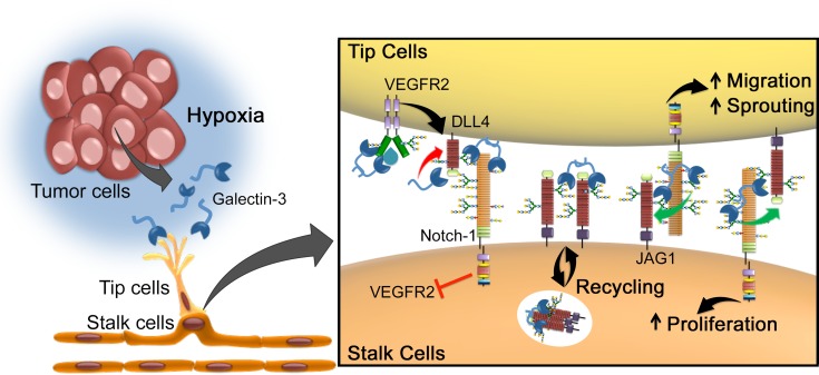
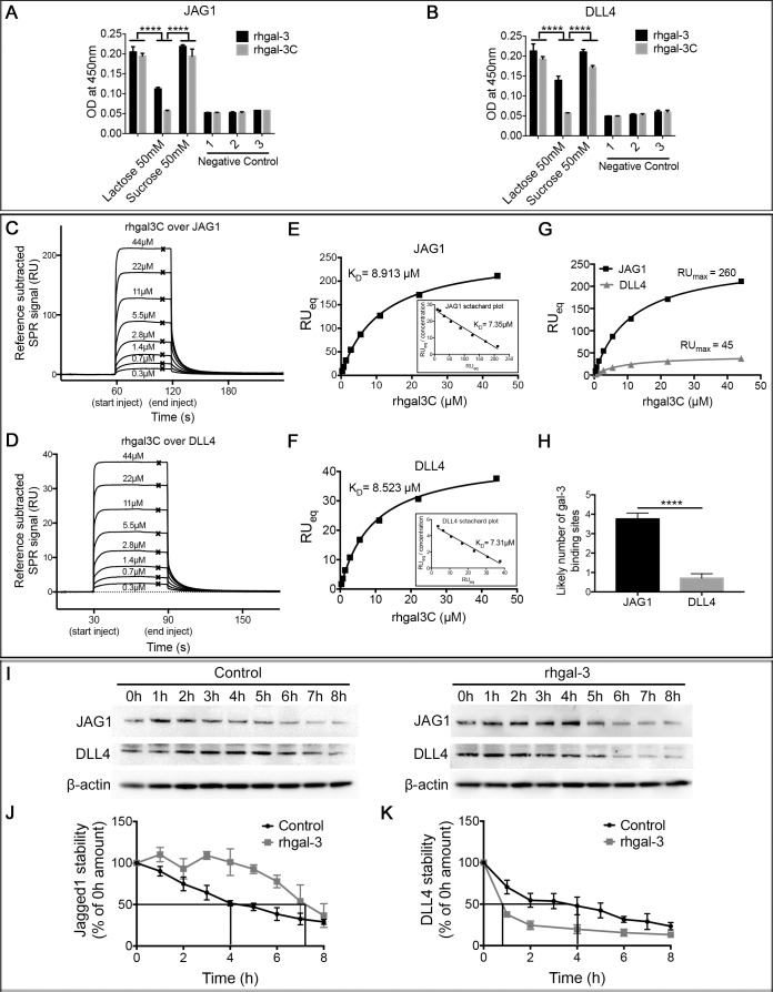
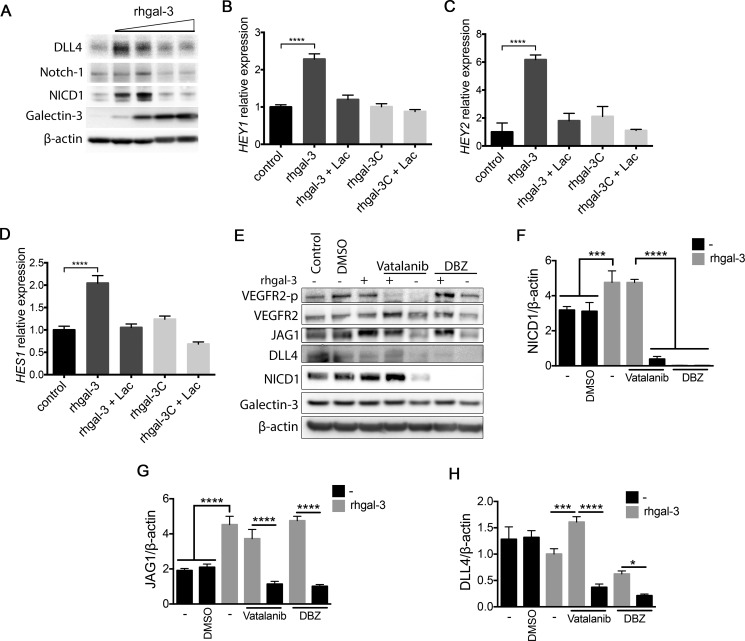
Angiogenesis is a coordinated process tightly regulated by the balance between Delta-like-4 (DLL4) and Jagged-1 (JAG1) in endothelial cells. Here we show that galectin-3 (gal-3), a glycan-binding protein secreted by cancer cells under hypoxic conditions, triggers sprouting angiogenesis, assisted by hypoxic changes in the glycosylation status of endothelial cells that enhance binding to gal-3. Galectin-3's proangiogenic functions were found to be predominantly dependent on the Notch ligand JAG1. Differential direct binding to JAG1 was shown by surface plasmon resonance assay. Upon binding to Notch ligands, gal-3 preferentially increased JAG1 protein half-life over DLL4 and preferentially activated JAG1/Notch-1 signaling in endothelial cells. JAG1 overexpression in Lewis lung carcinoma cells accelerated tumor growth in vivo, but this effect was prevented in Lgals3-/- mice. Our findings establish gal-3 as a molecular regulator of the JAG1/Notch-1 signaling pathway and have direct implications for the development of strategies aimed at controlling tumor angiogenesis.
血管生成是一个由内皮细胞中Delta样蛋白4(DLL4)和Jagged-1(JAG1)之间的平衡严格调控的协调过程。我们在此表明,半乳凝素-3(gal-3)是癌细胞在缺氧条件下分泌的一种聚糖结合蛋白,在内皮细胞糖基化状态的缺氧变化辅助下触发芽生血管生成,这种变化增强了与gal-3的结合。发现半乳凝素-3的促血管生成功能主要依赖于Notch配体JAG1。表面等离子体共振分析显示了与JAG1的差异直接结合。与Notch配体结合后,gal-3优先增加JAG1蛋白的半衰期而非DLL4,并优先激活内皮细胞中的JAG1/Notch-1信号通路。Lewis肺癌细胞中JAG1的过表达加速了体内肿瘤生长,但在Lgals3基因敲除小鼠中这种效应被阻止。我们的研究结果确立了gal-3作为JAG1/Notch-1信号通路的分子调节因子,并对旨在控制肿瘤血管生成的策略的开发具有直接意义。