Du Chen, Chai Ning-Li, Linghu En-Qiang, Li Hui-Kai, Sun Li-Hua, Jiang Lei, Wang Xiang-Dong, Tang Ping, Yang Jing
Chen Du, Ning-Li Chai, En-Qiang Linghu, Hui-Kai Li, Li-Hua Sun, Lei Jiang, Xiang-Dong Wang, Ping Tang, Jing Yang, Department of Gastroenterology and Hepatology, Chinese People's Liberation Army General Hospital, Beijing 100853, China.
World J Gastroenterol. 2017 May 7;23(17):3184-3192. doi: 10.3748/wjg.v23.i17.3184.
To evaluate the advantages of endoscopic ultrasound (EUS) in the assessment of detailed structures of pancreatic cystic neoplasms (PCNs) compared to computed tomography (CT) and magnetic resonance imaging (MRI).
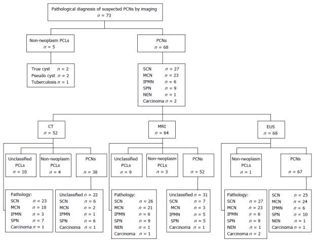
All patients with indeterminate PCNs underwent CT, MRI, and EUS. The detailed information, including size, number, the presence of a papilla/nodule, the presence of a septum, and the morphology of the pancreatic duct of PCNs were compared among the three imaging modalities. The size of each PCN was determined using the largest diameter measured. A cyst consisting of several small cysts was referred to as a mother-daughter cyst. Disagreement among the three imaging modalities regarding the total number of mother cysts resulted in the assumption that the correct number was the one in which the majority of imaging modalities indicated.
A total of 52 females and 16 males were evaluated. The median size of the cysts was 42.5 mm by EUS, 42.0 mm by CT and 38.0 mm by MRI; there was no significant difference in size as assessed among the three imaging techniques. The diagnostic sensitivity and ability of EUS to classify PCNs were 98.5% (67/68) and 92.6% (63/68), respectively. These percentages were higher than those of CT (73.1%, < 0.001; 17.1%, < 0.001) and MRI (81.3%, = 0.001; 20.3%, < 0.001). EUS was also able to better assess the number of daughter cysts in mother cysts than CT ( = 0.003); however, there was no significant difference between EUS and MRI in assessing mother-daughter cysts ( = 0.254). The papilla/nodule detection rate by EUS was 35.3% (24/68), much higher than those by CT (5.8%, 3/52) and MRI (6.3%, 4/64). The detection rate of the septum by EUS was 60.3% (41/68), which was higher than those by CT (34.6%, 18/52) and by MRI (46.9%, 30/64); the difference between EUS and CT was significant ( = 0.02). The rate of visualizing the pancreatic duct using EUS was 100%, whereas using CT and MRI it was less than 10%.
EUS helps visualize the detailed structures of PCNs and has many advantages over CT and MRI. EUS is valuable in the diagnosis and assessment of PCNs.
评估内镜超声(EUS)在评估胰腺囊性肿瘤(PCNs)详细结构方面相对于计算机断层扫描(CT)和磁共振成像(MRI)的优势。
所有PCNs诊断不明确的患者均接受了CT、MRI和EUS检查。比较了三种成像方式下PCNs的详细信息,包括大小、数量、乳头/结节的存在、隔膜的存在以及胰腺导管的形态。每个PCN的大小通过测量最大直径来确定。由几个小囊肿组成的囊肿被称为子母囊肿。三种成像方式在母囊肿总数上存在分歧,因此假设正确数量是大多数成像方式所显示的数量。
共评估了52名女性和16名男性。EUS测量的囊肿中位大小为42.5mm,CT为42.0mm,MRI为38.0mm;三种成像技术评估的大小无显著差异。EUS对PCNs的诊断敏感性和分类能力分别为98.5%(67/68)和92.6%(63/68)。这些百分比高于CT(73.1%,<0.001;17.1%,<0.001)和MRI(81.3%,=0.001;20.3%,<0.001)。EUS在评估母囊肿中子囊肿数量方面也比CT更好(=0.003);然而,EUS和MRI在评估子母囊肿方面无显著差异(=0.254)。EUS的乳头/结节检出率为35.3%(24/68),远高于CT(5.8%,3/52)和MRI(6.3%,4/64)。EUS的隔膜检出率为60.3%(41/68),高于CT(34.6%,18/52)和MRI(46.9%,30/64);EUS与CT之间的差异具有统计学意义(=0.02)。使用EUS观察胰腺导管的比率为100%,而使用CT和MRI时该比率低于10%。
EUS有助于观察PCNs的详细结构,与CT和MRI相比具有许多优势。EUS在PCNs的诊断和评估中具有重要价值。