Heard B J, Beveridge J E, Atarod M, O'Brien E J, Rolian C, Frank C B, Hart D A, Shrive N G
The McCaig Institute for Bone and Joint Health, University of Calgary, Calgary, AB, Canada.
Department of Comparative Biology and Experimental Medicine, Faculty of Veterinary Medicine, University of Calgary, Calgary, AB, Canada.
BMC Musculoskelet Disord. 2017 May 23;18(1):212. doi: 10.1186/s12891-017-1576-3.
Many patients who undergo anterior cruciate ligament (ACL) reconstructive surgery develop post-traumatic osteoarthritis (PTOA). ACL reconstructive surgery may not fully restore pre-injury joint biomechanics, thereby resulting in further joint damage and contributing to the development of PTOA. In an ovine model of idealized ACL reconstruction (ACL-R), it has been shown that signs of PTOA develop within surgical joints by 20 weeks post-surgery. The aim of the present study was to investigate whether altered kinematics contribute to early PTOA development within ACL-R joints of the ovine injury model by comparing the gait of these surgical animals to the gait of a stable normal control group, and an unstable injury group in which the ACL and medial collateral ligament (MCL) had been transected.
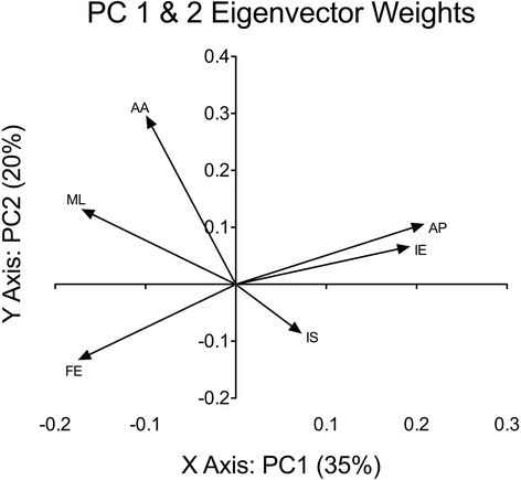
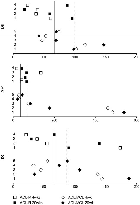
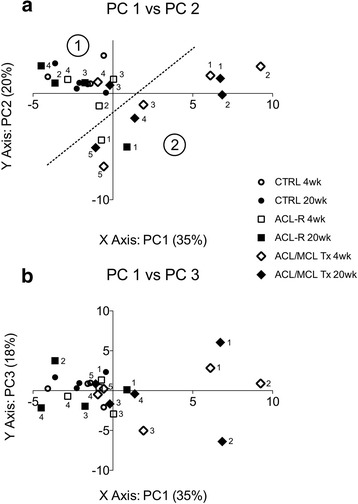
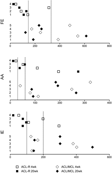
Fifteen skeletally mature female sheep were allocated evenly into 3 treatment groups: normal control, ACL-R, and ACL/MCL Tx (each group n = 5). Each animal's gait was recorded at baseline, 4 weeks post injury, and 20 weeks post injury. Principal component analysis (PCA) was used to identify the kinematic patterns that may be discriminant between treatment groups. Results from previous studies were referenced to present the amount of gross PTOA-like changes that occurred in the joints.
ACL-R and ACL/MCL transected (Tx) animals developed a similar amount of early PTOA-like changes within the surgical joints, but differed significantly in the amount of kinematic change present at 20 weeks post-surgery. We showed that the stifle joint kinematics of ACL/MCL Tx differed significantly from those of CTRL and the majority of ACL-R animals, while no significant differences in joint kinematic changes were found between ACL-R and CTRL animals.
These results suggest that the early PTOA-like changes reported in the ACL-R model cannot be attributed exclusively to post-surgical kinematic changes, and therefore biologic components in the post-injury environment must be contributing significantly to PTOA development.
许多接受前交叉韧带(ACL)重建手术的患者会发展为创伤后骨关节炎(PTOA)。ACL重建手术可能无法完全恢复损伤前的关节生物力学,从而导致进一步的关节损伤并促使PTOA的发展。在理想化ACL重建(ACL-R)的绵羊模型中,已表明在手术后20周内手术关节内会出现PTOA的迹象。本研究的目的是通过比较这些手术动物的步态与稳定的正常对照组以及ACL和内侧副韧带(MCL)已横断的不稳定损伤组的步态,来研究运动学改变是否有助于绵羊损伤模型中ACL-R关节内早期PTOA的发展。
将15只骨骼成熟的雌性绵羊平均分为3个治疗组:正常对照组、ACL-R组和ACL/MCL横断组(每组n = 5)。在基线、损伤后4周和损伤后20周记录每只动物的步态。主成分分析(PCA)用于识别可能区分治疗组的运动学模式。参考先前研究的结果来呈现关节中发生的大体PTOA样变化的量。
ACL-R组和ACL/MCL横断(Tx)组的动物在手术关节内出现了相似量的早期PTOA样变化,但在手术后20周时出现的运动学变化量有显著差异。我们表明,ACL/MCL Tx组的膝关节运动学与CTRL组和大多数ACL-R组动物有显著差异,而ACL-R组和CTRL组动物之间未发现关节运动学变化有显著差异。
这些结果表明,ACL-R模型中报告的早期PTOA样变化不能仅归因于手术后的运动学变化,因此损伤后环境中的生物学成分必定对PTOA的发展有重大影响。