Kim Dongjoon, Mecham Robert P, Trackman Philip C, Roy Sayon
Department of Medicine, Boston University School of Medicine, Boston, Massachusetts, United States 2Department of Ophthalmology, Boston University School of Medicine, Boston, Massachusetts, United States.
Department of Cell Biology and Physiology, Washington University School of Medicine, St. Louis, Missouri, United States.
Invest Ophthalmol Vis Sci. 2017 May 1;58(5):2725-2731. doi: 10.1167/iovs.16-21340.
To investigate the effect of reducing high glucose (HG)-induced lysyl oxidase (LOX) overexpression and increased activity on retinal endothelial cell apoptosis.
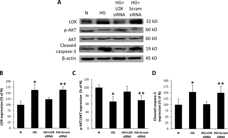
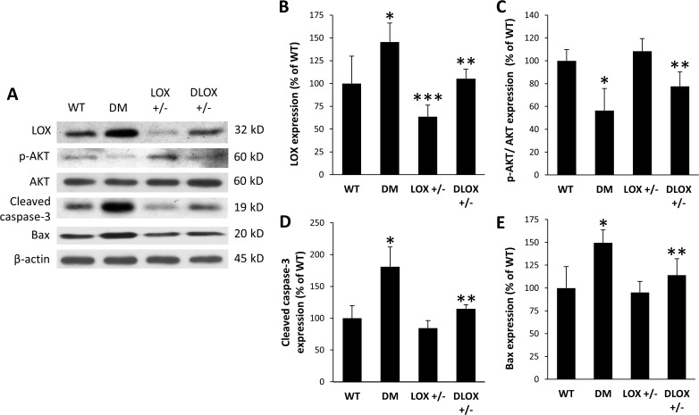
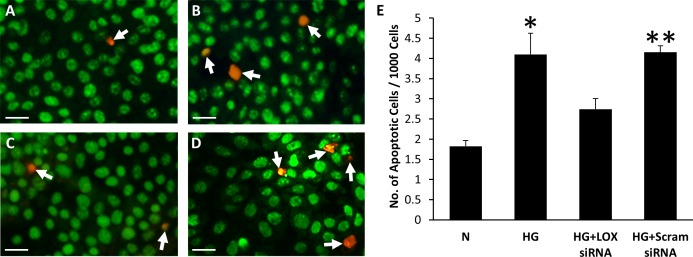
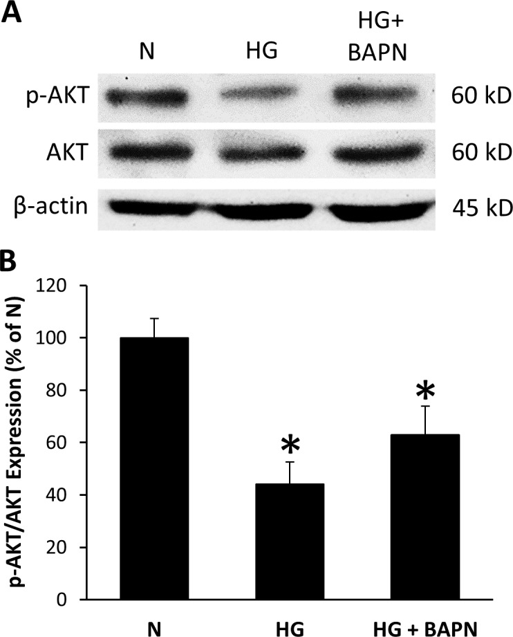
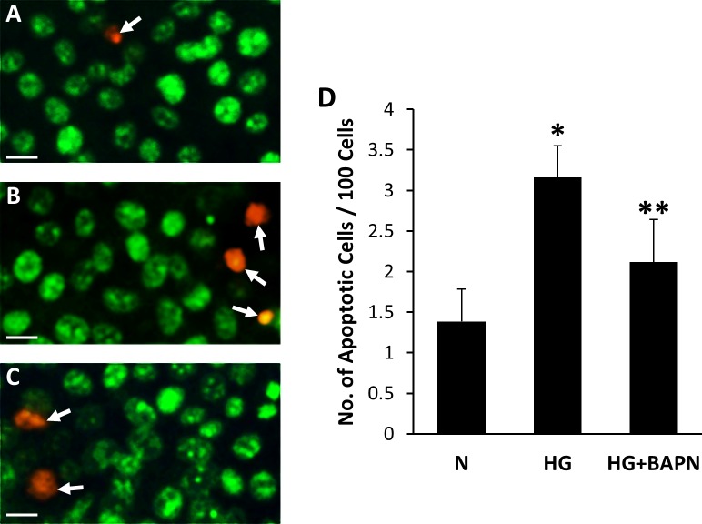
Rat retinal endothelial cells (RRECs) were grown in normal (N) or HG (30 mM glucose) medium for 7 days. In parallel, RRECs were grown in HG medium and transfected with LOX small interfering RNA (siRNA), scrambled siRNA as control, or exposed to β-aminopropionitrile (BAPN), a LOX inhibitor. LOX expression, AKT activation, and caspase-3 activity were determined by Western blot (WB) analysis and apoptosis by differential dye staining assay. Moreover, to determine whether diabetes-induced LOX overexpression alters AKT activation and promotes apoptosis, changes in LOX expression, AKT phosphorylation, caspase-3 activation, and Bax expression were assessed in retinas of streptozotocin (STZ)-induced diabetic mice and LOX heterozygous knockout (LOX+/-) mice.
WB analysis indicated significant LOX overexpression and reduced AKT activation under HG condition in RRECs. Interestingly, when cells grown in HG were transfected with LOX siRNA or exposed to BAPN, the number of apoptotic cells was significantly decreased concomitant with increased AKT phosphorylation. Diabetic mouse retinas exhibited LOX overexpression, decreased AKT phosphorylation, and increased Bax and caspase-3 activation compared to values in nondiabetic mice. In LOX+/- mice, reduced LOX levels were observed with increased AKT activity, and reduced Bax and caspase-3 activity. Furthermore, decreased levels of LOX in the LOX+/- mice was protective against diabetes-induced apoptosis.
Findings from this study indicate that preventing LOX overexpression may be protective against HG-induced apoptosis in retinal vascular cells associated with diabetic retinopathy.
研究降低高糖(HG)诱导的赖氨酰氧化酶(LOX)过表达及活性增加对视网膜内皮细胞凋亡的影响。
大鼠视网膜内皮细胞(RRECs)在正常(N)或HG(30 mM葡萄糖)培养基中培养7天。同时,RRECs在HG培养基中培养,并用LOX小干扰RNA(siRNA)转染,以乱序siRNA作为对照,或用LOX抑制剂β-氨基丙腈(BAPN)处理。通过蛋白质免疫印迹(WB)分析测定LOX表达、AKT激活和半胱天冬酶-3活性,通过差异染料染色法测定细胞凋亡。此外,为了确定糖尿病诱导的LOX过表达是否会改变AKT激活并促进凋亡,在链脲佐菌素(STZ)诱导的糖尿病小鼠和LOX杂合敲除(LOX+/-)小鼠的视网膜中评估了LOX表达、AKT磷酸化、半胱天冬酶-3激活和Bax表达的变化。
WB分析表明,在HG条件下,RRECs中LOX显著过表达,AKT激活降低。有趣的是,当在HG中生长的细胞用LOX siRNA转染或暴露于BAPN时,凋亡细胞数量显著减少,同时AKT磷酸化增加。与非糖尿病小鼠相比,糖尿病小鼠视网膜表现出LOX过表达、AKT磷酸化降低以及Bax和半胱天冬酶-3激活增加。在LOX+/-小鼠中,观察到LOX水平降低,AKT活性增加,Bax和半胱天冬酶-3活性降低。此外,LOX+/-小鼠中LOX水平降低对糖尿病诱导的凋亡具有保护作用。
本研究结果表明,预防LOX过表达可能对糖尿病视网膜病变相关的视网膜血管细胞中HG诱导的凋亡具有保护作用。