By Samantha, Xu Junzhong, Box Bailey A, Bagnato Francesca R, Smith Seth A
Department of Biomedical Engineering, Vanderbilt University, Nashville, TN, USA; Vanderbilt University Institute of Imaging Science, Vanderbilt University Medical Center, Nashville, TN, USA.
Department of Biomedical Engineering, Vanderbilt University, Nashville, TN, USA; Vanderbilt University Institute of Imaging Science, Vanderbilt University Medical Center, Nashville, TN, USA; Department of Radiology and Radiological Sciences, Vanderbilt University Medical Center, Nashville, TN, USA.
Neuroimage Clin. 2017 May 17;15:333-342. doi: 10.1016/j.nicl.2017.05.010. eCollection 2017.
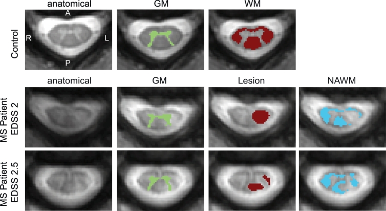
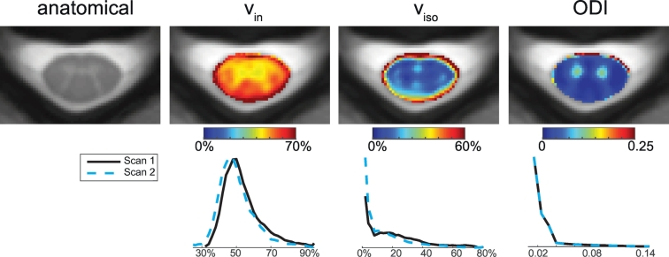
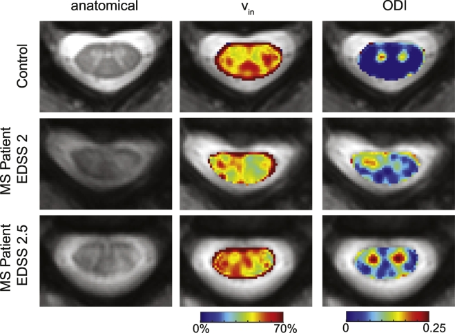
There is a need to develop imaging methods sensitive to axonal injury in multiple sclerosis (MS), given the prominent impact of axonal pathology on disability and outcome. Advanced multi-compartmental diffusion models offer novel indices sensitive to white matter microstructure. One such model, neurite orientation dispersion and density imaging (NODDI), is sensitive to neurite morphology, providing indices of apparent volume fractions of axons (v), isotropic water (v) and the dispersion of fibers about a central axis (orientation dispersion index, ODI). NODDI has yet to be studied for its sensitivity to spinal cord pathology. Here, we investigate the feasibility and utility of NODDI in the cervical spinal cord of MS patients.
NODDI was applied in the cervical spinal cord in a cohort of 8 controls and 6 MS patients. Statistical analyses were performed to test the sensitivity of NODDI-derived indices to pathology in MS (both lesion and normal appearing white matter NAWM). Diffusion kurtosis imaging (DKI) and diffusion tensor imaging (DTI) analysis were also performed to compare with NODDI.
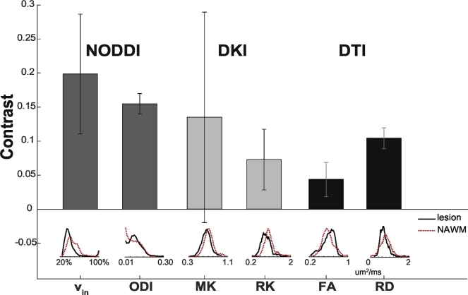
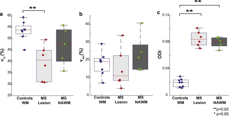
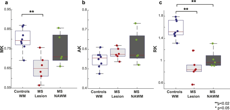
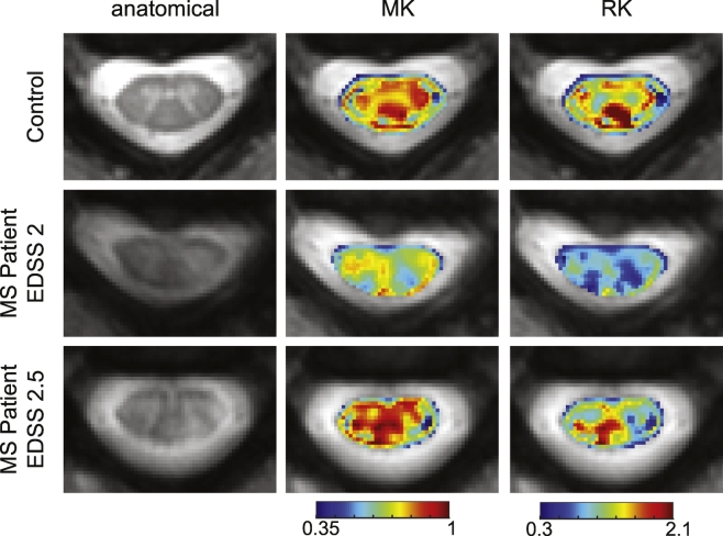
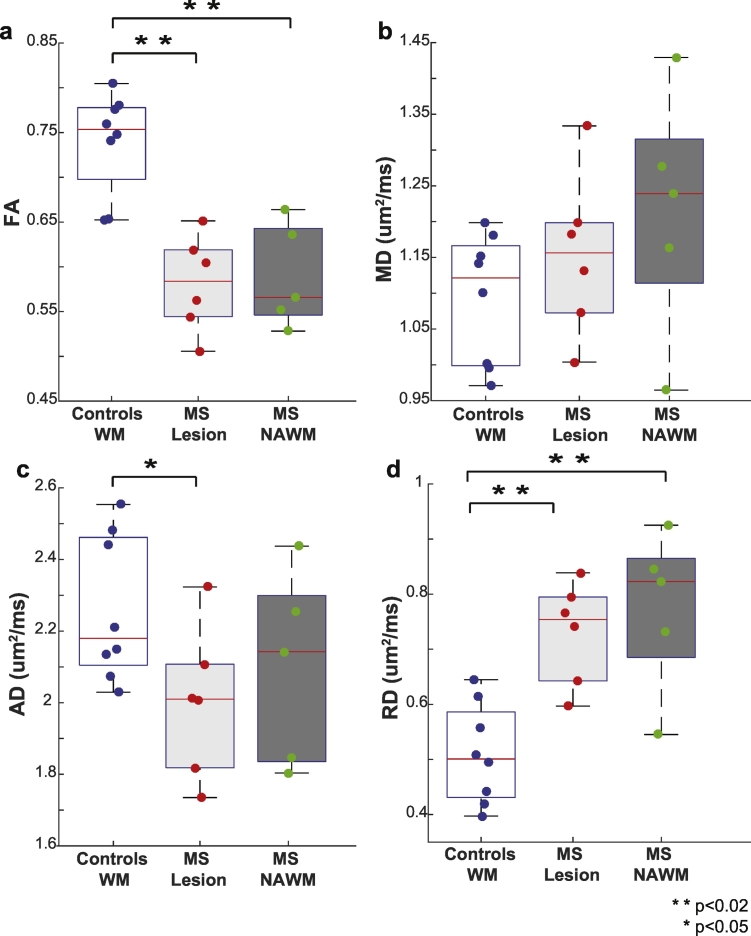
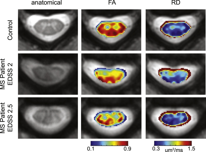
A decrease in NODDI-derived v was observed at the site of the lesion ( < 0.01), whereas a global increase in ODI was seen throughout white matter ( < 0.001). DKI-derived mean kurtosis (MK) and radial kurtosis (RK) and DTI-derived fractional anisotropy (FA) and radial diffusivity (RD) were all significantly different in MS patients ( < 0.02), however NODDI provided higher contrast between NAWM and lesion in all MS patients.
NODDI provides unique contrast that is not available with DKI or DTI, enabling improved characterization of the spinal cord in MS.
鉴于轴突病理对多发性硬化症(MS)患者残疾程度和预后的显著影响,有必要开发对MS轴突损伤敏感的成像方法。先进的多室扩散模型提供了对白质微观结构敏感的新指标。其中一种模型,即神经突方向离散度与密度成像(NODDI),对神经突形态敏感,可提供轴突表观体积分数(v)、各向同性水(v)以及纤维围绕中心轴的离散度(方向离散度指数,ODI)等指标。NODDI对脊髓病理的敏感性尚未得到研究。在此,我们研究NODDI在MS患者颈脊髓中的可行性和实用性。
对8名对照者和6名MS患者的颈脊髓进行NODDI检查。进行统计分析以测试NODDI衍生指标对MS病理(病变和正常白质NAWM)的敏感性。还进行了扩散峰度成像(DKI)和扩散张量成像(DTI)分析以与NODDI进行比较。
在病变部位观察到NODDI衍生的v降低(<0.01),而在整个白质中ODI整体升高(<0.001)。MS患者的DKI衍生的平均峰度(MK)和径向峰度(RK)以及DTI衍生的分数各向异性(FA)和径向扩散率(RD)均有显著差异(<0.02),然而在所有MS患者中,NODDI在NAWM和病变之间提供了更高的对比度。
NODDI提供了DKI或DTI无法提供的独特对比度,能够更好地表征MS患者的脊髓。