• 文献检索
  • 文档翻译
  • 深度研究
  • 学术资讯
  • Suppr Zotero 插件Zotero 插件
  • 邀请有礼
  • 套餐&价格
  • 历史记录
应用&插件
Suppr Zotero 插件Zotero 插件浏览器插件Mac 客户端Windows 客户端微信小程序
定价
高级版会员购买积分包购买API积分包
服务
文献检索文档翻译深度研究API 文档MCP 服务
关于我们
关于 Suppr公司介绍联系我们用户协议隐私条款
关注我们

Suppr 超能文献

核心技术专利:CN118964589B侵权必究
粤ICP备2023148730 号-1Suppr @ 2026

文献检索

告别复杂PubMed语法,用中文像聊天一样搜索,搜遍4000万医学文献。AI智能推荐,让科研检索更轻松。

立即免费搜索

文件翻译

保留排版,准确专业,支持PDF/Word/PPT等文件格式,支持 12+语言互译。

免费翻译文档

深度研究

AI帮你快速写综述,25分钟生成高质量综述,智能提取关键信息,辅助科研写作。

立即免费体验

组成型活性Alk5在肝细胞中的特异性表达会加剧硫代乙酰胺诱导的小鼠肝损伤。

Hepatocyte-specific expression of constitutively active Alk5 exacerbates thioacetamide-induced liver injury in mice.

作者信息

Kongphat Wanthita, Pudgerd Arnon, Sridurongrit Somyoth

机构信息

Graduate Program of Toxicology, Faculty of Science, Mahidol University, Bangkok, Thailand.

Department of Anatomy, Faculty of Science, Mahidol University, Bangkok, Thailand.

出版信息

Heliyon. 2017 May 19;3(5):e00305. doi: 10.1016/j.heliyon.2017.e00305. eCollection 2017 May.

DOI:10.1016/j.heliyon.2017.e00305
PMID:28560358
原文链接:https://pmc.ncbi.nlm.nih.gov/articles/PMC5440359/
Abstract

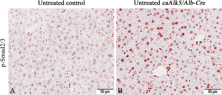
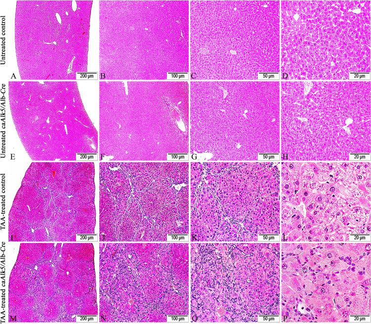
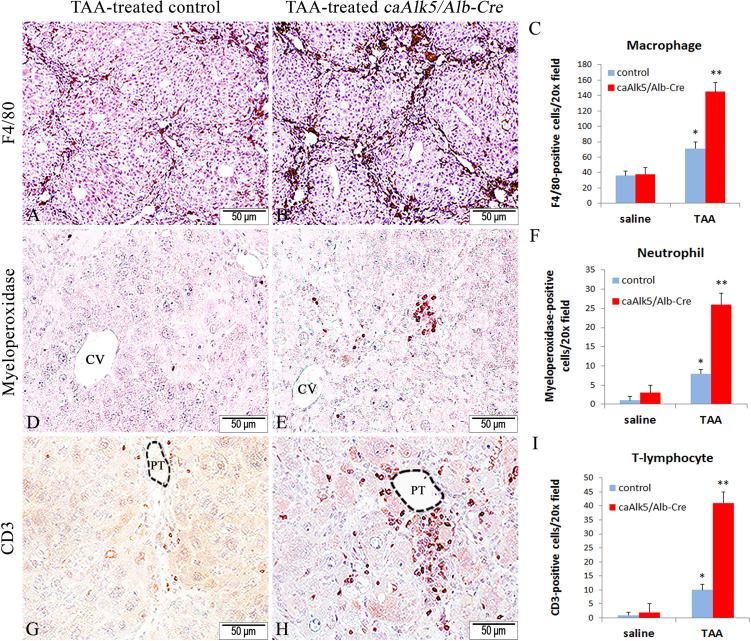
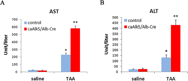
While Transforming growth factor-βs (Tgf-βs) have been known to play an important role in liver fibrosis through an activation of Hepatic Stellate Cells (HSC), their fibrotic role on hepatocytes in liver damage has not been addressed thoroughly. To shed more light on the hepatocyte-specific role of Tgf-β signaling during liver fibrosis, we generated transgenic mice expressing constitutively active Tgf-β type I receptor Alk5 under the control of albumin promoter. Uninjured mice with increased Tgf-β/Alk5 signaling in hepatocytes ( mice) did not show characteristics related to hepatocyte death, fibrosis and inflammation. When subjected to thioacetamide (TAA) treatment, mice exhibited more severe liver injury, when compared to control littermates. After TAA administration for 12 weeks, an increase in pathological changes was evident in livers, with higher number of infiltrating cells in the portal and periportal area. Immunohistochemistry for F4/80, myeloperoxidase and CD3 showed that there was an increased accumulation of macrophages, neutrophils and T-lymphocytes, respectively, in livers. Coincidently, we observed an exacerbated liver damage as seen by increases in serum aminotransferase level and number of apoptotic hepatocytes in mice. Sirius staining of collagen demonstrated that the fibrotic response was worsened in mice. The enhanced fibrosis in mutant livers was associated with marked production of α-SMA-positive myofibroblast. Hepatic expression of genes indicative of HSC activation was greater in mice. In conclusion, our data indicated that elevation of Tgf-β signaling via Alk5 in hepatocytes is not sufficient to induce liver pathology but plays an important role in amplifying TAA-induced liver damage.

摘要

虽然已知转化生长因子-β(Tgf-β)通过激活肝星状细胞(HSC)在肝纤维化中发挥重要作用,但其在肝损伤中对肝细胞的纤维化作用尚未得到充分研究。为了更深入了解肝纤维化过程中Tgf-β信号在肝细胞中的特异性作用,我们构建了在白蛋白启动子控制下组成型表达活性Tgf-β I型受体Alk5的转基因小鼠。肝细胞中Tgf-β/Alk5信号增加的未受伤小鼠(小鼠)未表现出与肝细胞死亡、纤维化和炎症相关的特征。与对照同窝小鼠相比,用硫代乙酰胺(TAA)处理时,小鼠表现出更严重的肝损伤。给予TAA 12周后,小鼠肝脏的病理变化明显增加,门脉和门脉周围区域的浸润细胞数量更多。F4/80、髓过氧化物酶和CD3的免疫组织化学显示,小鼠肝脏中巨噬细胞、中性粒细胞和T淋巴细胞的积累分别增加。巧合的是,我们观察到小鼠血清转氨酶水平升高和凋亡肝细胞数量增加,肝损伤加剧。天狼星胶原染色显示,小鼠的纤维化反应恶化。突变肝脏中增强的纤维化与α-SMA阳性肌成纤维细胞的大量产生有关。小鼠肝脏中指示HSC激活的基因表达更高。总之,我们的数据表明,肝细胞中通过Alk-5升高的Tgf-β信号不足以诱导肝脏病变,但在放大TAA诱导的肝损伤中起重要作用。

https://cdn.ncbi.nlm.nih.gov/pmc/blobs/1d28/5440359/8b7f71fc553d/gr10.jpg
https://cdn.ncbi.nlm.nih.gov/pmc/blobs/1d28/5440359/68e89b9e6621/gr1.jpg
https://cdn.ncbi.nlm.nih.gov/pmc/blobs/1d28/5440359/f49ce0f7778b/gr2.jpg
https://cdn.ncbi.nlm.nih.gov/pmc/blobs/1d28/5440359/716337d7cdd4/gr3.jpg
https://cdn.ncbi.nlm.nih.gov/pmc/blobs/1d28/5440359/acea95b9e37f/gr4.jpg
https://cdn.ncbi.nlm.nih.gov/pmc/blobs/1d28/5440359/dce49be31ebd/gr5.jpg
https://cdn.ncbi.nlm.nih.gov/pmc/blobs/1d28/5440359/b57a22d4d935/gr6.jpg
https://cdn.ncbi.nlm.nih.gov/pmc/blobs/1d28/5440359/2da786093189/gr7.jpg
https://cdn.ncbi.nlm.nih.gov/pmc/blobs/1d28/5440359/a642c9e13aed/gr8.jpg
https://cdn.ncbi.nlm.nih.gov/pmc/blobs/1d28/5440359/e4feed153ed0/gr9.jpg
https://cdn.ncbi.nlm.nih.gov/pmc/blobs/1d28/5440359/8b7f71fc553d/gr10.jpg

相似文献

1
Hepatocyte-specific expression of constitutively active Alk5 exacerbates thioacetamide-induced liver injury in mice.组成型活性Alk5在肝细胞中的特异性表达会加剧硫代乙酰胺诱导的小鼠肝损伤。
Heliyon. 2017 May 19;3(5):e00305. doi: 10.1016/j.heliyon.2017.e00305. eCollection 2017 May.
2
Cell-specific PPARγ deficiency establishes anti-inflammatory and anti-fibrogenic properties for this nuclear receptor in non-parenchymal liver cells.细胞特异性过氧化物酶体增殖物激活受体 γ 缺失赋予非实质细胞中该核受体抗炎和抗纤维化特性。
J Hepatol. 2013 Nov;59(5):1045-53. doi: 10.1016/j.jhep.2013.06.023. Epub 2013 Jul 2.
3
Cooperation of liver cells in health and disease.健康与疾病状态下肝细胞的协作。
Adv Anat Embryol Cell Biol. 2001;161:III-XIII, 1-151. doi: 10.1007/978-3-642-56553-3.
4
Selective deletion of hepatocyte platelet-derived growth factor receptor α and development of liver fibrosis in mice.选择性敲除肝细胞血小板衍生生长因子受体α导致小鼠肝纤维化的发生。
Cell Commun Signal. 2018 Dec 3;16(1):93. doi: 10.1186/s12964-018-0306-2.
5
Essential role of suppressor of cytokine signaling 1 (SOCS1) in hepatocytes and macrophages in the regulation of liver fibrosis.细胞因子信号转导抑制因子 1(SOCS1)在肝细胞和巨噬细胞中对肝纤维化调节的重要作用。
Cytokine. 2019 Dec;124:154501. doi: 10.1016/j.cyto.2018.07.032. Epub 2018 Aug 8.
6
Hepatocyte-specific deletion of ARNT (aryl hydrocarbon Receptor Nuclear Translocator) results in altered fibrotic gene expression in the thioacetamide model of liver injury.肝细胞特异性缺失芳烃受体核转运蛋白(ARNT)会导致硫代乙酰胺肝损伤模型中纤维化基因表达发生改变。
PLoS One. 2015 Mar 26;10(3):e0121650. doi: 10.1371/journal.pone.0121650. eCollection 2015.
7
TGF-β Type I Receptor Signaling in Melanoma Liver Metastases Increases Metastatic Outgrowth.TGF-β 型 I 受体信号在黑色素瘤肝转移中增加转移生长。
Int J Mol Sci. 2023 May 12;24(10):8676. doi: 10.3390/ijms24108676.
8
Hepatocyte-specific METTL3 ablation by Alb-iCre mice (GPT), but not by Alb-Cre mice (JAX), resulted in acute liver failure (ALF) and postnatal lethality.Alb-iCre 小鼠(GPT)而非 Alb-Cre 小鼠(JAX)特异性敲除肝细胞中的 METTL3 导致急性肝衰竭(ALF)和出生后致死。
Aging (Albany NY). 2024 Apr 22;16(8):7217-7248. doi: 10.18632/aging.205753.
9
Detrimental effects of nicotine on thioacetamide-induced liver injury in mice.尼古丁对硫代乙酰胺诱导的小鼠肝损伤的有害影响。
Toxicol Mech Methods. 2017 Sep;27(7):501-510. doi: 10.1080/15376516.2017.1323256. Epub 2017 May 16.
10
[Mechanism of hepatocyte mitochondrial NDUFA13 deficiency-induced liver fibrogenesis: the role of abnormal hepatic stellate cell activation].[肝细胞线粒体NDUFA13缺乏诱导肝纤维化的机制:肝星状细胞异常激活的作用]
Nan Fang Yi Ke Da Xue Xue Bao. 2021 Apr 20;41(4):529-535. doi: 10.12122/j.issn.1673-4254.2021.04.07.

引用本文的文献

1
Macrophage expression of constitutively active alleviates hepatic injury in a mouse model of concanavalin A-induced autoimmune hepatitis.组成型活性巨噬细胞的表达减轻了刀豆蛋白A诱导的自身免疫性肝炎小鼠模型中的肝损伤。
Heliyon. 2025 Feb 13;11(4):e42691. doi: 10.1016/j.heliyon.2025.e42691. eCollection 2025 Feb 28.
2
Liver regeneration during the associating liver partition and portal vein ligation for staged hepatectomy procedure in is positively modulated by stem cells.在联合肝脏分隔和门静脉结扎分期肝切除术过程中,肝脏再生受到干细胞的正向调节。
Oncol Lett. 2018 May;15(5):6309-6321. doi: 10.3892/ol.2018.8108. Epub 2018 Feb 22.

本文引用的文献

1
Galectin-3, histone deacetylases, and Hedgehog signaling: Possible convergent targets in schistosomiasis-induced liver fibrosis.半乳糖凝集素-3、组蛋白去乙酰化酶与刺猬信号通路:血吸虫病诱导肝纤维化中可能的共同作用靶点
PLoS Negl Trop Dis. 2017 Feb 23;11(2):e0005137. doi: 10.1371/journal.pntd.0005137. eCollection 2017 Feb.
2
Inhibition of Notch Signaling Attenuates Schistosomiasis Hepatic Fibrosis via Blocking Macrophage M2 Polarization.Notch信号通路的抑制通过阻断巨噬细胞M2极化减轻血吸虫病肝纤维化。
PLoS One. 2016 Nov 22;11(11):e0166808. doi: 10.1371/journal.pone.0166808. eCollection 2016.
3
Activation of the miR-34a/SIRT1/p53 Signaling Pathway Contributes to the Progress of Liver Fibrosis via Inducing Apoptosis in Hepatocytes but Not in HSCs.
miR-34a/SIRT1/p53信号通路的激活通过诱导肝细胞而非肝星状细胞凋亡促进肝纤维化进展。
PLoS One. 2016 Jul 7;11(7):e0158657. doi: 10.1371/journal.pone.0158657. eCollection 2016.
4
Liver regeneration - mechanisms and models to clinical application.肝脏再生 - 从机制到模型到临床应用。
Nat Rev Gastroenterol Hepatol. 2016 Aug;13(8):473-85. doi: 10.1038/nrgastro.2016.97. Epub 2016 Jun 29.
5
TGF-β: the master regulator of fibrosis.TGF-β:纤维化的主调控因子。
Nat Rev Nephrol. 2016 Jun;12(6):325-38. doi: 10.1038/nrneph.2016.48. Epub 2016 Apr 25.
6
TGF-β signalling and liver disease.转化生长因子-β信号传导与肝脏疾病
FEBS J. 2016 Jun;283(12):2219-32. doi: 10.1111/febs.13665. Epub 2016 Feb 22.
7
Dectin-1 Regulates Hepatic Fibrosis and Hepatocarcinogenesis by Suppressing TLR4 Signaling Pathways.Dectin-1通过抑制TLR4信号通路调控肝纤维化和肝癌发生。
Cell Rep. 2015 Dec 1;13(9):1909-1921. doi: 10.1016/j.celrep.2015.10.058. Epub 2015 Nov 19.
8
Epithelial Transforming Growth Factor-β Signaling Does Not Contribute to Liver Fibrosis but Protects Mice From Cholangiocarcinoma.上皮转化生长因子-β信号通路对肝纤维化无作用,但可保护小鼠免受胆管癌侵害。
Gastroenterology. 2016 Mar;150(3):720-33. doi: 10.1053/j.gastro.2015.11.039. Epub 2015 Nov 26.
9
Structural determinants of Smad function in TGF-β signaling.转化生长因子-β信号通路中Smad功能的结构决定因素
Trends Biochem Sci. 2015 Jun;40(6):296-308. doi: 10.1016/j.tibs.2015.03.012. Epub 2015 Apr 29.
10
A novel mouse model of depletion of stellate cells clarifies their role in ischemia/reperfusion- and endotoxin-induced acute liver injury.一种新型的星状细胞耗竭小鼠模型阐明了它们在缺血/再灌注和内毒素诱导的急性肝损伤中的作用。
J Hepatol. 2014 Feb;60(2):298-305. doi: 10.1016/j.jhep.2013.09.013. Epub 2013 Sep 20.