Liu Jianlin, Dai Juan, Wang Yansong, Lai Siyu, Wang Suwen
Department of Stomatology, Shenzhen Hospital of Guangzhou University of Chinese Medicine, Shenzhen, Guangdong 518033, P.R. China.
Exp Ther Med. 2017 May;13(5):2325-2331. doi: 10.3892/etm.2017.4234. Epub 2017 Mar 17.
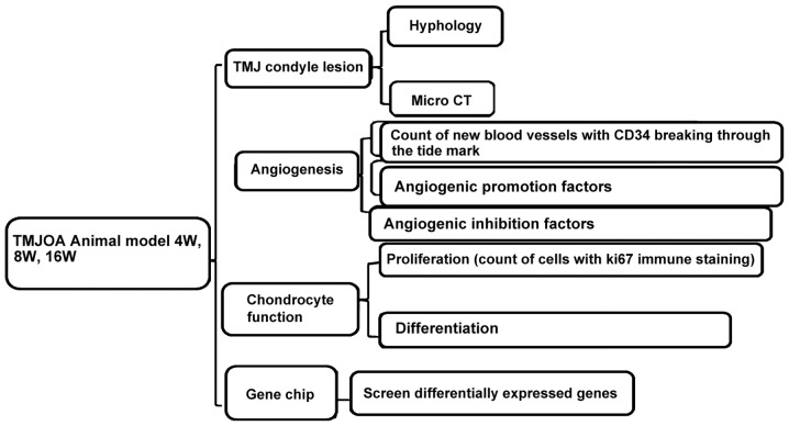
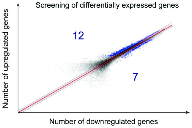
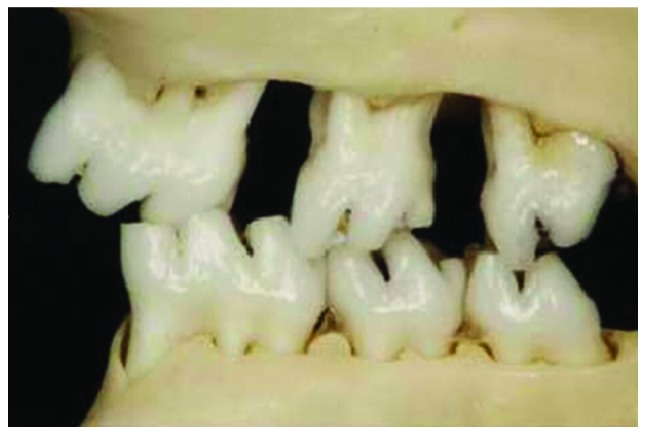
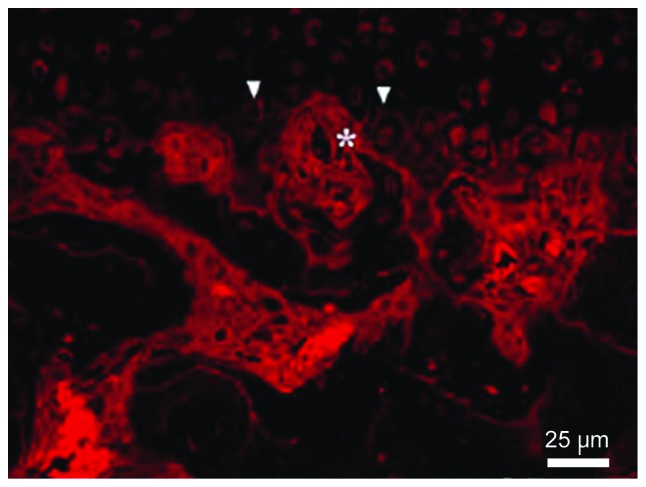
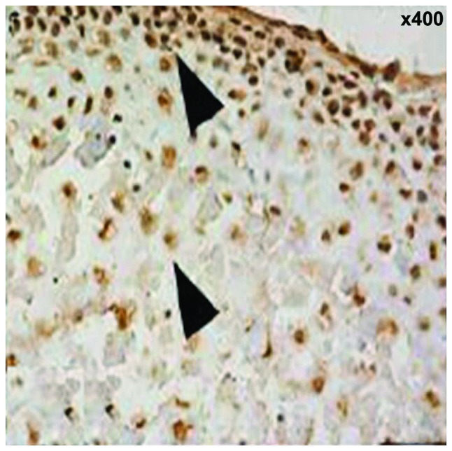
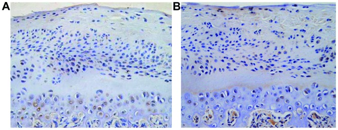
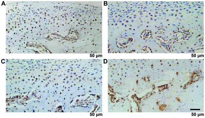
We studied the significance of new blood vessels in the pathogenesis of temporomandibular joint osteoarthritis (TMJOA). Fifteen 8-week-old female Sprague-Dawley rats were selected to establish TMJOA models of gradually induced occlusal disorders. Five rats were sacrificed at 4, 8 and 16 weeks, and histological exam was conducted along with micro-computed tomography observation on the condyle specimen. The distribution and number of new blood vessels breaking were observed through the tidemark through CD34 immunofluorescence staining. The proliferation of chondrocytes were detected through Ki67 immunohistochemical staining, and the differentiation functions of chondrocytes were observed through PTHrP and IHH immunohistochemical staining. The degradation functions of cartilage matrix were observed through matrix metalloproteinase (MMP)-9 immunohistochemical staining to detect the expression of vascular growth promotion and inhibition factors with vascular endothelial growth factor (VEGF), CTGF and CHM-1 immunohistochemical staining and screen differentially expressed genes through gene chip analysis method. It was found that the condyle tissue full thickness, fiber layer thickness and calcified cartilage layer thickness were significantly increased with time (P<0.05). Bone mineral density, trabecular thickness and Tb.Sp were also increased significantly with time, BS/BV and trabecular number were decreased significantly with time (P<0.05). The new blood vessels reached the deep layer of calcified cartilage until the tide line was broken and non-calcified cartilage was invaded. The number of vessels were increased significantly with time (P<0.05). Ki67, PTHrP and IHH-positive rates were increased significantly (P<0.05). MMP-9, VEGF, CTGF and CHM-1 were increased significantly (P<0.05). VEGF, CTGF and CHM-1 mRNA were upregulated differentially with the expressed genes. In conclusion, the new blood vessels may be important in the pathogenesis of TMJOA.
我们研究了新生血管在颞下颌关节骨关节炎(TMJOA)发病机制中的意义。选取15只8周龄雌性Sprague-Dawley大鼠,建立逐渐诱导咬合紊乱的TMJOA模型。分别于4周、8周和16周处死5只大鼠,对髁突标本进行组织学检查及显微计算机断层扫描观察。通过CD34免疫荧光染色观察穿过潮线的新生血管的分布及数量。通过Ki67免疫组织化学染色检测软骨细胞的增殖情况,通过甲状旁腺激素相关蛋白(PTHrP)和印度刺猬蛋白(IHH)免疫组织化学染色观察软骨细胞的分化功能。通过基质金属蛋白酶(MMP)-9免疫组织化学染色观察软骨基质的降解功能,用血管内皮生长因子(VEGF)、结缔组织生长因子(CTGF)和CHM-1免疫组织化学染色检测血管生长促进和抑制因子的表达,并采用基因芯片分析方法筛选差异表达基因。结果发现,髁突组织全层厚度、纤维层厚度和钙化软骨层厚度随时间显著增加(P<0.05)。骨密度、骨小梁厚度和骨小梁间距也随时间显著增加,骨表面积与骨体积比和骨小梁数量随时间显著减少(P<0.05)。新生血管到达钙化软骨深层,直至潮线断裂,非钙化软骨受到侵犯。血管数量随时间显著增加(P<0.05)。Ki67、PTHrP和IHH阳性率显著升高(P<0.05)。MMP-9、VEGF、CTGF和CHM-1显著升高(P<0.05)。VEGF、CTGF和CHM-1 mRNA与表达基因呈差异上调。综上所述,新生血管可能在TMJOA的发病机制中起重要作用。