Eikendal Anouk L M, den Ruijter Hester M, Haaring Cees, Saam Tobias, van der Geest Rob J, Westenberg Jos J M, Bots Michiel L, Hoefer Imo E, Leiner Tim
Department of Radiology (E01.132), University Medical Center Utrecht, Heidelberglaan 100, 3584 CX, Utrecht, The Netherlands.
Laboratory of Experimental Cardiology, University Medical Center Utrecht, Heidelberglaan 100, 3584 CX, Utrecht, The Netherlands.
MAGMA. 2018 Feb;31(1):173-182. doi: 10.1007/s10334-017-0626-z. Epub 2017 May 31.
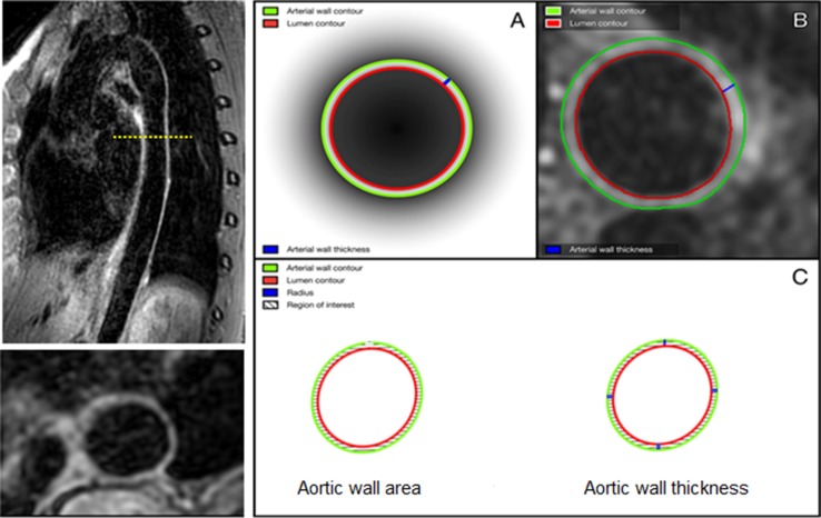
More detailed evaluation of atherosclerosis and its key determinants in young individuals is warranted to improve knowledge on the pathophysiology of its development and progression. This study evaluated associations of magnetic resonance imaging (MRI)-derived aortic wall area, wall thickness, and pulse wave velocity (PWV) with cardiovascular risk factors in asymptomatic, young adults.
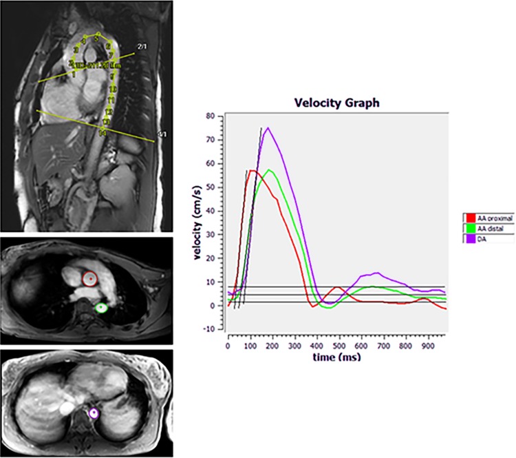
In 124 adults (age: 25-35 years) from the general population-based Atherosclerosis Monitoring and Biomarker Measurements in the Young study, demography, anthropometry, and blood samples were collected. The studied MRI-parameters were measured using a 3.0T MRI system. Relations between cardiovascular risk factors and aortic characteristics were assessed using multivariable linear regression analyses.
Mean age was 31.8 years, 47.6% was male. Aortic wall area was positively associated with age [β = 0.01, (95% confidence interval (CI) 2.00 × 10, 0.02), p = 0.01] and BMI [β = 0.01, (0.01, 0.02), p = 0.003] and negatively associated with sex (reference: men) [β = -0.06, (-0.11, -0.01), p = 0.02]. Natural logarithm transformed (ln) aortic wall thickness was positively associated with BMI [β = 0.01, (1.00 × 10, 0.02), p = 0.02]. Ln aortic PWV was positively associated with 10 mmHg increment of SBP [β = 0.06, (0.03, 0.09), p < 0.001] and DBP [β = 0.06, (0.02, 0.09), p = 0.006]. No relations were observed for smoking and lipids.
Already in early adulthood, aortic wall geometry and stiffness vary by age, sex, BMI, and blood pressure.
有必要对年轻人的动脉粥样硬化及其关键决定因素进行更详细的评估,以增进对其发生和发展病理生理学的了解。本研究评估了磁共振成像(MRI)得出的主动脉壁面积、壁厚和脉搏波速度(PWV)与无症状年轻成年人心血管危险因素之间的关联。
在基于一般人群的“年轻人动脉粥样硬化监测和生物标志物测量”研究中的124名成年人(年龄:25 - 35岁)中,收集了人口统计学、人体测量学和血液样本。使用3.0T MRI系统测量研究的MRI参数。使用多变量线性回归分析评估心血管危险因素与主动脉特征之间的关系。
平均年龄为31.8岁,男性占47.6%。主动脉壁面积与年龄呈正相关[β = 0.01,(95%置信区间(CI)2.00×10,0.02),p = 0.01]和BMI呈正相关[β = 0.01,(0.01,0.02),p = 0.003],与性别(参照:男性)呈负相关[β = -0.06,(-0.11,-0.01),p = 0.02]。自然对数转换后的(ln)主动脉壁厚与BMI呈正相关[β = 0.01,(1.00×10,0.02),p = 0.02]。Ln主动脉PWV与收缩压(SBP)升高10 mmHg呈正相关[β = 0.06,(0.03,0.09),p < 0.001],与舒张压(DBP)呈正相关[β = 0.06,(0.02,0.09),p = 0.006]。未观察到吸烟和血脂之间的关系。
在成年早期,主动脉壁几何形状和硬度就因年龄、性别、BMI和血压而有所不同。