ElSheikh Mona, Arani Arvin, Perry Avital, Boeve Bradley F, Meyer Fredric B, Savica Rodolfo, Ehman Richard L, Huston John
1 Department of Radiology, Mayo Clinic, 200 First St SW, Rochester, MN 55905.
2 Department of Neurologic Surgery, Mayo Clinic, Rochester, MN.
AJR Am J Roentgenol. 2017 Aug;209(2):403-408. doi: 10.2214/AJR.16.17455. Epub 2017 Jun 1.
The purpose of this study was to investigate age-corrected brain MR elastography (MRE) findings in four dementia cohorts (Alzheimer disease, dementia with Lewy bodies, frontotemporal dementia, and normal pressure hydrocephalus) and determine the potential use as a differentiating biomarker in dementia subtypes.
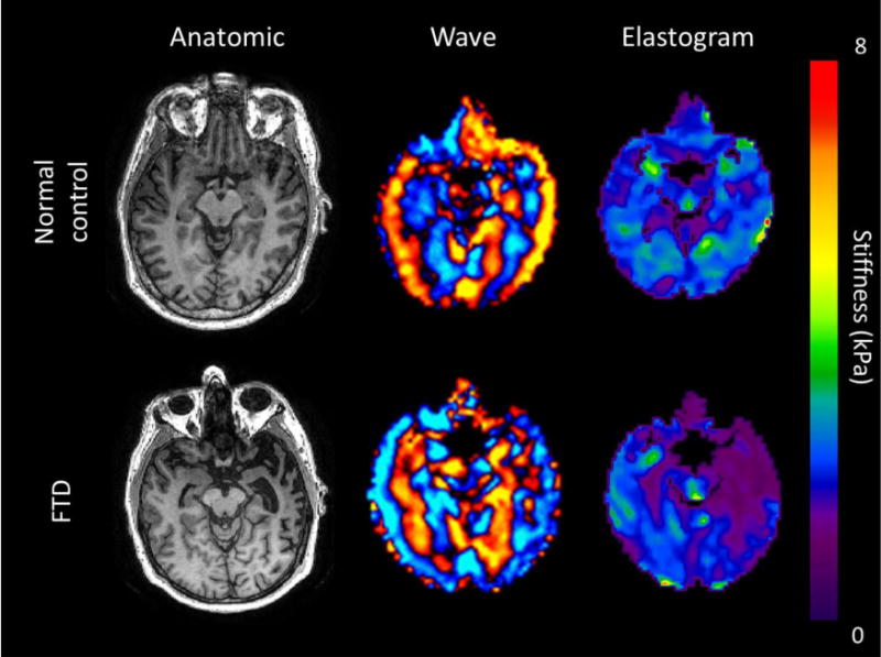
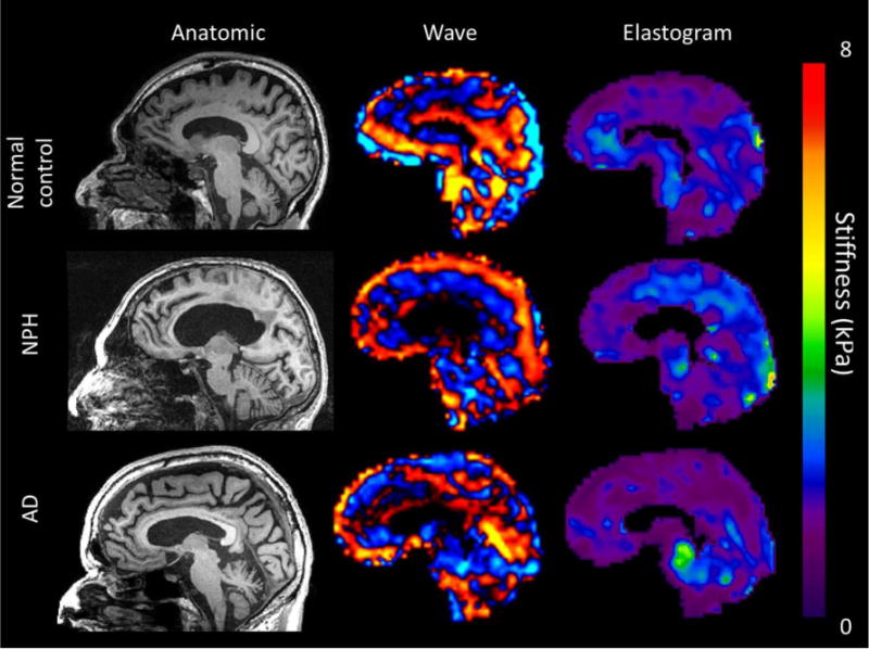
Institutional review board approval and written informed consent were obtained to perform MRE on 84 subjects: 20 patients with normal pressure hydrocephalus, eight with Alzheimer disease, five with dementia with Lewy bodies, five with frontotemporal dementia, and 46 cognitively normal control subjects. Shear waves of 60-Hz vibration frequency were transmitted into the brain using a pillowlike passive driver, and brain stiffness was determined in eight different regions (cerebrum, frontal, occipital, parietal, temporal, deep gray matter-white matter, sensorimotor cortex, and cerebellum). All stiffness values were age-corrected and compared with control subjects. The Wilcoxon rank sum test and linear regression were used for statistical analysis.
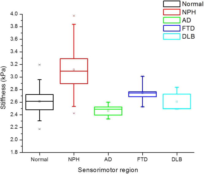
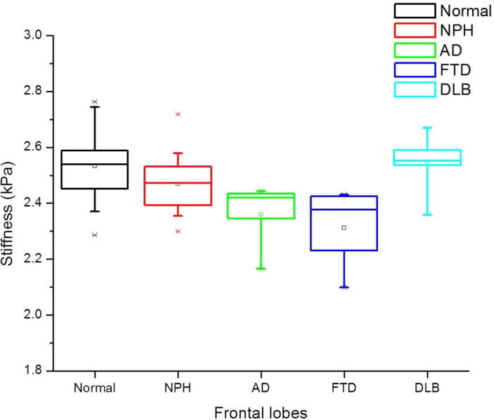
Regional stiffness patterns unique to each dementing disorder were observed. Patients with Alzheimer disease and frontotemporal dementia showed decreased cerebral stiffness (p = 0.001 and p = 0.002, respectively) with regional softening of the frontal and temporal lobes. Patients with Alzheimer disease additionally showed parietal lobe and sensorimotor region softening (p = 0.039 and p = 0.018, respectively). Patients with normal pressure hydrocephalus showed stiffening of the parietal, occipital, and sensorimotor regions (p = 0.007, p < 0.001, and p < 0.0001, respectively). Patients with dementia with Lewy bodies did not show significant stiffness changes in any of the regions.
Quantitative MRE of changes in brain viscoelastic structure shows unique regional brain stiffness patterns between common dementia subtypes.
本研究旨在调查四个痴呆队列(阿尔茨海默病、路易体痴呆、额颞叶痴呆和正常压力脑积水)中经年龄校正的脑磁共振弹性成像(MRE)结果,并确定其作为痴呆亚型鉴别生物标志物的潜在用途。
获得机构审查委员会批准并取得书面知情同意后,对84名受试者进行MRE检查,其中包括20例正常压力脑积水患者、8例阿尔茨海默病患者、5例路易体痴呆患者、5例额颞叶痴呆患者以及46名认知正常的对照受试者。使用类似枕头的被动驱动器将60赫兹振动频率的剪切波传入大脑,并在八个不同区域(大脑、额叶、枕叶、顶叶、颞叶、深部灰质-白质、感觉运动皮层和小脑)测定脑硬度。所有硬度值均进行了年龄校正,并与对照受试者进行比较。采用Wilcoxon秩和检验和线性回归进行统计分析。
观察到每种痴呆症特有的区域硬度模式。阿尔茨海默病和额颞叶痴呆患者的大脑硬度降低(分别为p = 0.001和p = 0.002),额叶和颞叶出现区域软化。阿尔茨海默病患者还表现出顶叶和感觉运动区域软化(分别为p = 0.039和p = 0.018)。正常压力脑积水患者的顶叶、枕叶和感觉运动区域变硬(分别为p = 0.007、p < 0.001和p < 0.0001)。路易体痴呆患者在任何区域均未显示出明显的硬度变化。
脑粘弹性结构变化的定量MRE显示,常见痴呆亚型之间存在独特的区域脑硬度模式。