• 文献检索
  • 文档翻译
  • 深度研究
  • 学术资讯
  • Suppr Zotero 插件Zotero 插件
  • 邀请有礼
  • 套餐&价格
  • 历史记录
应用&插件
Suppr Zotero 插件Zotero 插件浏览器插件Mac 客户端Windows 客户端微信小程序
定价
高级版会员购买积分包购买API积分包
服务
文献检索文档翻译深度研究API 文档MCP 服务
关于我们
关于 Suppr公司介绍联系我们用户协议隐私条款
关注我们

Suppr 超能文献

核心技术专利:CN118964589B侵权必究
粤ICP备2023148730 号-1Suppr @ 2026

文献检索

告别复杂PubMed语法,用中文像聊天一样搜索,搜遍4000万医学文献。AI智能推荐,让科研检索更轻松。

立即免费搜索

文件翻译

保留排版,准确专业,支持PDF/Word/PPT等文件格式,支持 12+语言互译。

免费翻译文档

深度研究

AI帮你快速写综述,25分钟生成高质量综述,智能提取关键信息,辅助科研写作。

立即免费体验

人角膜基质偏振干涉测量分析的干涉图

Interference figures of polarimetric interferometry analysis of the human corneal stroma.

作者信息

Mastropasqua Rodolfo, Nubile Mario, Salgari Niccolò, Lanzini Manuela, Calienno Roberta, Mattei Peter A, Sborgia Alessandra, Agnifili Luca

机构信息

Ophthalmology Unit, Department of Neurological, Neuropsychological, Morphological and Movement Sciences, University of Verona, Verona, Italy.

National Centre of High Technology (CNAT) in Ophthalmology of University "G. d'Annunzio", Chieti-Pescara, Italy.

出版信息

PLoS One. 2017 Jun 1;12(6):e0178397. doi: 10.1371/journal.pone.0178397. eCollection 2017.

DOI:10.1371/journal.pone.0178397
PMID:28570631
原文链接:https://pmc.ncbi.nlm.nih.gov/articles/PMC5453517/
Abstract

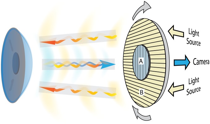
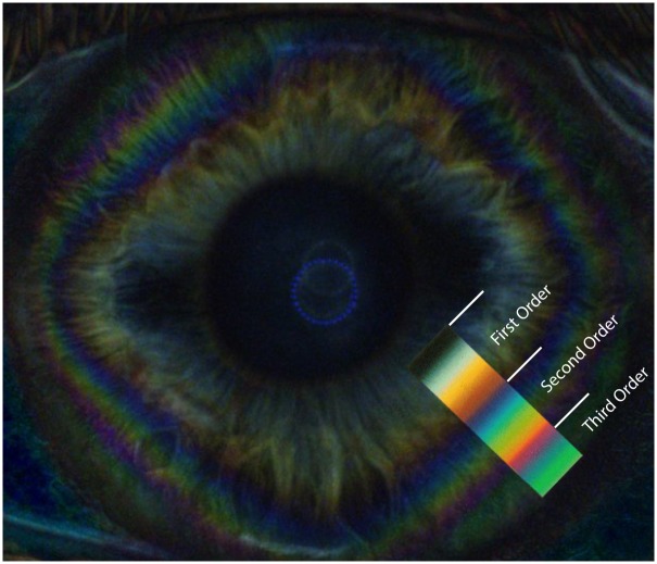
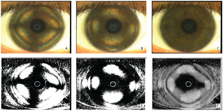
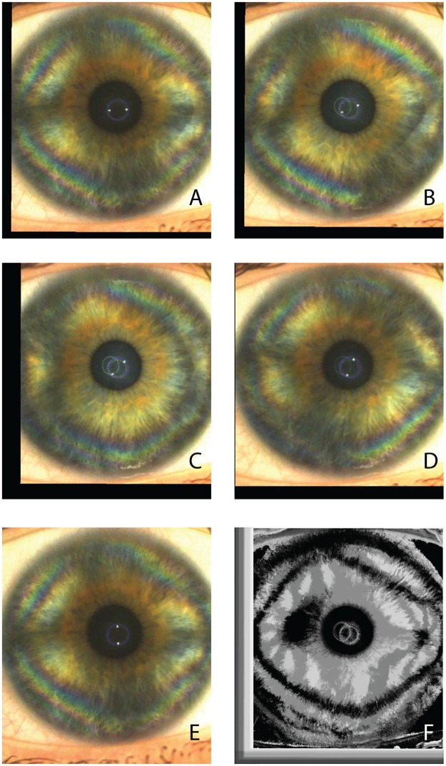
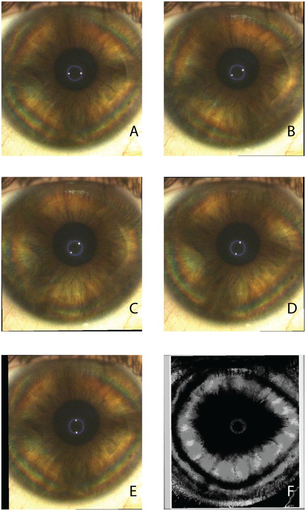
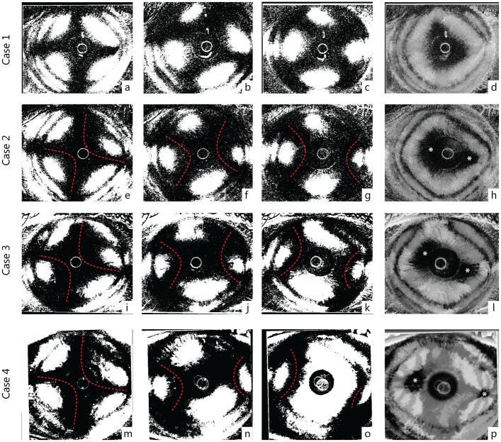
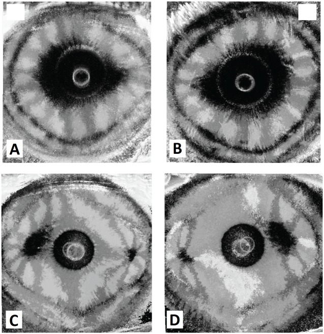
A rotating polarimetric 90°-cross linear-filter interferometry system was used to detect the morphological characteristics and features of interference patterns produced in in-vivo corneal stroma in healthy human corneas of 23 subjects. The characteristic corneal isogyres presenting with an evident cross-shaped pattern, grossly aligned with the fixation axis, were observed in all patients with centers within the pupillary dark area, impeding the exact determination of the center point. During the rotational scan in 78.3% of the eyes the cross-shaped pattern of the isogyre gradually separated to form two distinct hyperbolic arcs in opposite quadrants, reaching their maximal separation at 45 degrees with respect to angle of cross-shaped pattern formation. The corneal cross and hyperbolic-pattern repeated every 90° throughout the 360° rotational scan. While the interpretation of the isogyres presents particular difficulties, two summary parameters can be extracted for each cornea: the presence/orientation of a single or two dark areas in post-processed images and isochromes. However, the development of dedicated software for semi-quantitative analysis of these parameters and enantiomorphism may become available in the near future. The possible application of polarimetric interferometry in the field of both corneal pathologies and corneal surgery may be of great interest for clinical purposes.

摘要

使用旋转偏振90°交叉线性滤光干涉测量系统检测23名受试者健康人角膜体内角膜基质中产生的干涉图样的形态特征。在所有患者中均观察到特征性的角膜等视线呈现明显的十字形图案,大致与注视轴对齐,其中心位于瞳孔暗区内,妨碍了中心点的精确测定。在78.3%的眼睛旋转扫描过程中,等视线的十字形图案逐渐分开,在相对象限形成两个明显的双曲线弧,相对于十字形图案形成角度在45度时达到最大分离。在360°旋转扫描过程中,角膜十字和双曲线图案每90°重复一次。虽然对等视线的解释存在特殊困难,但可以为每个角膜提取两个汇总参数:后处理图像和等色线中单个或两个暗区的存在/方向。然而,用于这些参数和对映体半定量分析的专用软件可能在不久的将来问世。偏振干涉测量在角膜病理学和角膜手术领域的可能应用可能对临床目的具有极大的意义。

https://cdn.ncbi.nlm.nih.gov/pmc/blobs/d4d8/5453517/2c23840f1ab2/pone.0178397.g007.jpg
https://cdn.ncbi.nlm.nih.gov/pmc/blobs/d4d8/5453517/1995dab13c62/pone.0178397.g001.jpg
https://cdn.ncbi.nlm.nih.gov/pmc/blobs/d4d8/5453517/c65b20a57029/pone.0178397.g002.jpg
https://cdn.ncbi.nlm.nih.gov/pmc/blobs/d4d8/5453517/548ff1d07dd1/pone.0178397.g003.jpg
https://cdn.ncbi.nlm.nih.gov/pmc/blobs/d4d8/5453517/728c50ab84df/pone.0178397.g004.jpg
https://cdn.ncbi.nlm.nih.gov/pmc/blobs/d4d8/5453517/6175b2de5814/pone.0178397.g005.jpg
https://cdn.ncbi.nlm.nih.gov/pmc/blobs/d4d8/5453517/0fc935b946c7/pone.0178397.g006.jpg
https://cdn.ncbi.nlm.nih.gov/pmc/blobs/d4d8/5453517/2c23840f1ab2/pone.0178397.g007.jpg

相似文献

1
Interference figures of polarimetric interferometry analysis of the human corneal stroma.人角膜基质偏振干涉测量分析的干涉图
PLoS One. 2017 Jun 1;12(6):e0178397. doi: 10.1371/journal.pone.0178397. eCollection 2017.
2
Polarimetric Interferometry for Assessment of Corneal Stromal Lamellae Orientation.用于评估角膜基质板层方向的偏振干涉测量法。
Cornea. 2016 Apr;35(4):519-22. doi: 10.1097/ICO.0000000000000776.
3
Subclinical keratoconus detection by pattern analysis of corneal and epithelial thickness maps with optical coherence tomography.通过光学相干断层扫描对角膜和上皮厚度图进行模式分析检测亚临床圆锥角膜
J Cataract Refract Surg. 2016 Feb;42(2):284-95. doi: 10.1016/j.jcrs.2015.09.021.
4
[Confocal microscopy of the corneal after photorefractive keratectomy with the excimer laser].[准分子激光屈光性角膜切削术后角膜的共聚焦显微镜检查]
Klin Monbl Augenheilkd. 1997 Sep;211(3):159-67. doi: 10.1055/s-2008-1035116.
5
Posterior linear stromal haze formation after simultaneous photorefractive keratectomy followed by corneal collagen cross-linking.准分子激光原位角膜磨镶术联合角膜胶原交联术后的后部线性基质混浊形成
Invest Ophthalmol Vis Sci. 2010 Oct;51(10):5030-3. doi: 10.1167/iovs.09-5105. Epub 2010 May 12.
6
Estimating corneal surface topography in videokeratoscopy in the presence of strong signal interference.在存在强信号干扰的情况下估算视频角膜地形图中的角膜表面地形。
IEEE Trans Biomed Eng. 2008 Oct;55(10):2381-7. doi: 10.1109/TBME.2008.923766.
7
Determination of corneal volume from anterior topography and topographic pachymetry: application to healthy and keratoconic eyes.从前表面地形图和地形角膜测厚术测定角膜体积:在健康眼和圆锥角膜眼中的应用。
Ophthalmic Physiol Opt. 2009 Nov;29(6):652-60. doi: 10.1111/j.1475-1313.2009.00642.x.
8
[An analysis about the safe range of thickness of the residual corneal stroma bed after LASIK].[关于准分子激光原位角膜磨镶术(LASIK)后残余角膜基质床厚度安全范围的分析]
Zhonghua Yan Ke Za Zhi. 2004 Nov;40(11):741-4.
9
Collagen fibril orientation in the human corneal stroma and its implication in keratoconus.人角膜基质中胶原纤维的取向及其在圆锥角膜中的意义。
Invest Ophthalmol Vis Sci. 1997 Jan;38(1):121-9.
10
In vivo confocal microscopy findings in a patient with posterior amorphous corneal dystrophy.一名后部无定形角膜营养不良患者的体内共焦显微镜检查结果
Clin Exp Ophthalmol. 2007 Jan-Feb;35(1):99-102. doi: 10.1111/j.1442-9071.2007.01426.x.

引用本文的文献

1
Collagen imaging reveals synergistic effects of sutures and host-donor misalignment on topographical irregularities in penetrating keratoplasty.胶原成像揭示了缝线和供体-宿主错位对穿透性角膜移植术中地形不规则性的协同作用。
PLoS One. 2024 Aug 8;19(8):e0308204. doi: 10.1371/journal.pone.0308204. eCollection 2024.
2
An integrated model of the human cornea as a linear biaxial birefringent medium.人眼角膜作为一种线性双轴双折射介质的综合模型。
Sci Rep. 2024 Mar 1;14(1):5077. doi: 10.1038/s41598-024-55800-4.
3
Birefringent properties of the cornea measured by a Mueller type polarimeter in healthy adults and children.

本文引用的文献

1
Polarimetric Interferometry for Assessment of Corneal Stromal Lamellae Orientation.用于评估角膜基质板层方向的偏振干涉测量法。
Cornea. 2016 Apr;35(4):519-22. doi: 10.1097/ICO.0000000000000776.
2
Corneal enantiomorphism in normal and keratoconic eyes.正常眼与圆锥角膜眼中的角膜对称性
J Refract Surg. 2014 Aug;30(8):542-7. doi: 10.3928/1081597X-20140711-07.
3
NIH Image to ImageJ: 25 years of image analysis.NIH 图像到 ImageJ:25 年的图像分析。
通过穆勒型偏振计测量健康成人和儿童角膜的双折射特性。
Biomed Opt Express. 2021 Dec 1;12(12):7872-7885. doi: 10.1364/BOE.440274.
4
Coherent poly propagation materials with 3-dimensional photonic control over visible light.具有三维光子控制可见光的相干多聚物传播材料。
PLoS One. 2019 Oct 17;14(10):e0223715. doi: 10.1371/journal.pone.0223715. eCollection 2019.
Nat Methods. 2012 Jul;9(7):671-5. doi: 10.1038/nmeth.2089.
4
In vivo structural imaging of the cornea by polarization-resolved second harmonic microscopy.通过偏振分辨二次谐波显微镜对角膜进行体内结构成像。
Biomed Opt Express. 2012 Jan 1;3(1):1-15. doi: 10.1364/BOE.3.000001. Epub 2011 Dec 1.
5
Three-dimensional analysis of collagen lamellae in the anterior stroma of the human cornea visualized by second harmonic generation imaging microscopy.利用二次谐波产生成像显微镜观察人眼角膜前基质中胶原层的三维分析。
Invest Ophthalmol Vis Sci. 2011 Feb 16;52(2):911-5. doi: 10.1167/iovs.10-5657.
6
Effect of granule size on the interference colors of starch in polarized light.颗粒大小对淀粉在偏振光下干涉色的影响。
Biotech Histochem. 2009 Dec;84(6):329-36. doi: 10.3109/10520290903081369.
7
3D collagen orientation study of the human cornea using X-ray diffraction and femtosecond laser technology.利用X射线衍射和飞秒激光技术对人角膜进行三维胶原取向研究。
Invest Ophthalmol Vis Sci. 2009 Nov;50(11):5159-64. doi: 10.1167/iovs.09-3669. Epub 2009 Jun 10.
8
Investigation of the polarization optics of the living human cornea and lens with purkinje images.利用浦肯野图像对活体人眼角膜和晶状体的偏振光学进行研究。
Appl Opt. 1998 Oct 1;37(28):6845-51. doi: 10.1364/ao.37.006845.
9
Optical anisotropy of the human cornea determined with a polarizing microscope.用偏光显微镜测定人角膜的光学各向异性。
Appl Opt. 2007 Dec 1;46(34):8351-7. doi: 10.1364/ao.46.008351.
10
Circular polarization biomicroscopy: a method for determining human corneal stromal lamellar organization in vivo.圆偏振生物显微镜检查法:一种用于在体内确定人角膜基质层状结构的方法。
Ophthalmic Physiol Opt. 2007 May;27(3):256-64. doi: 10.1111/j.1475-1313.2007.00482.x.