Adachi Kazuhide, Hasegawa Mitsuhiro, Hirose Yuichi
Department of Neurosurgery, School of Medicine, Fujita Health University.
Neurol Med Chir (Tokyo). 2017 Oct 15;57(10):505-512. doi: 10.2176/nmc.ra.2016-0336. Epub 2017 Jun 5.
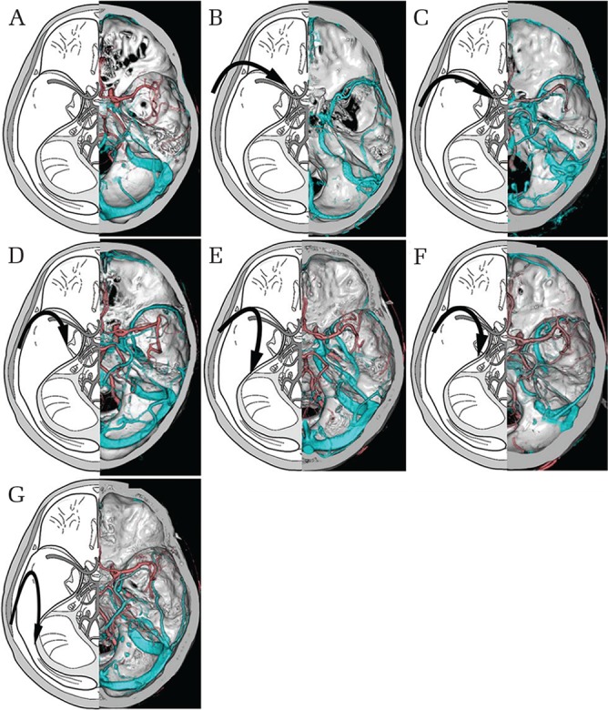
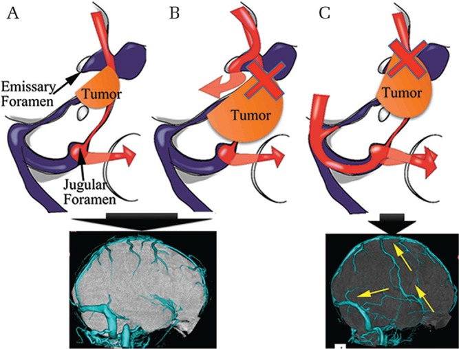
The evaluation of venous drainage patterns prior to surgery for skull base meningioma is important owing to their deep location and the vulnerability of surrounding vascular structures. In recent years, the microsurgical skull base approach has matured as a surgical technique, making it an important option for reducing complications related to skull base meningioma surgery. In addition, knowledge of the venous anatomy can prevent venous drainage route disturbance and potentially life-threatening complications. Hence, this topic review aimed to provide an overview of normal venous anatomy as it relates to the microsurgical skull base approach, discuss known changes in venous drainage routes that are associated with the progression of skull base meningioma and the selection of an appropriate operative approach with the highest likelihood of preserving venous drainage structures.
由于颅底脑膜瘤位置深且周围血管结构脆弱,术前评估静脉引流模式很重要。近年来,显微外科颅底入路作为一种手术技术已成熟,成为减少颅底脑膜瘤手术相关并发症的重要选择。此外,了解静脉解剖结构可防止静脉引流途径紊乱及潜在的危及生命的并发症。因此,本专题综述旨在概述与显微外科颅底入路相关的正常静脉解剖结构,讨论与颅底脑膜瘤进展相关的已知静脉引流途径变化,以及选择最有可能保留静脉引流结构的合适手术入路。