Karami V, Zabihzadeh M
Medical Physics Student (MSc), Department of Medical Physics, School of Medicine, Ahvaz Jundishapur University of Medical Sciences, Ahvaz, Iran.
Assistant Professor (PhD), Department of Medical Physics, School of Medicine, Ahvaz Jundishapur University of Medical Sciences, Ahvaz, Iran.
J Biomed Phys Eng. 2017 Jun 1;7(2):101-106. eCollection 2017 Jun.
Collimating the primary beam to the area of diagnostic interest (ADI) has been strongly recommended as an effective method to reduce patient's radiation dose and to improve image quality during radiology practice. Lack or inadequate collimation results in excessive radiation dose to patients and deterioration image quality.
To assess the quality of beam collimation during lumbar spine radiography at two general hospitals in Ahvaz, Iran.
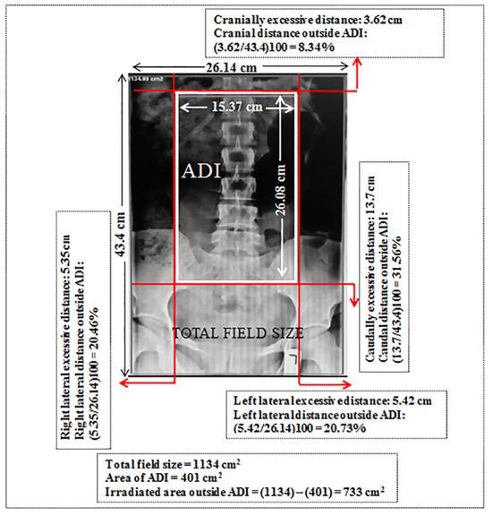
We retrospectively reviewed 830 digital antero-posterior (AP) lumbar spine radiographs in term of beam collimation. For each radiograph, the distance between current and optimal collimation was calculated (in cm). The area of ADI and total field size for each radiograph were also calculated (in cm).
The total mean ADI and irradiated region outside ADI for each radiograph were estimated 360 and 454 cm, respectively. The total irradiated region outside ADI was 1.26 times more than ADI. In contrast to cranial regions outside ADI, caudal regions were more commonly included inside the primary beam (12% vs. 24.4%; P-value <0.005). At least in 62% of radiographs evaluated, ovaries were included in the primary beam.
Radiographers should make considerable effort to limit the primary beam to the ADI to reduce patient's exposure and to increase image quality.
强烈推荐将主射线束准直至诊断感兴趣区域(ADI),作为在放射学实践中降低患者辐射剂量和提高图像质量的有效方法。准直不足或缺乏会导致患者接受过量辐射剂量,并使图像质量下降。
评估伊朗阿瓦士两家综合医院腰椎摄影时的射线束准直质量。
我们回顾性地分析了830张数字化腰椎前后位(AP)X线片的射线束准直情况。对于每张X线片,计算当前准直与最佳准直之间的距离(单位:厘米)。还计算了每张X线片的ADI面积和总视野大小(单位:厘米)。
每张X线片的平均ADI总面积和ADI外的受照区域分别估计为360平方厘米和454平方厘米。ADI外的总受照区域比ADI大1.26倍。与ADI外的头侧区域相比,尾侧区域更常被包括在主射线束内(12%对24.4%;P值<0.005)。在至少62%的评估X线片中,卵巢被包括在主射线束内。
放射技师应做出相当大的努力,将主射线束限制在ADI范围内,以减少患者的辐射暴露并提高图像质量。