Fujino Yoko, Minamizaki Tomoko, Yoshioka Hirotaka, Okada Mitsugi, Yoshiko Yuji
Department of Special Care Dentistry, Hiroshima University Graduate School of Biomedical and Health Sciences, Hiroshima University, Hiroshima, Japan.
Department of Calcified Tissue Biology, Hiroshima University Institute of Biomedical and Health Sciences, Hiroshima University, Hiroshima, Japan.
Bone Rep. 2016 Sep 29;5:280-285. doi: 10.1016/j.bonr.2016.09.004. eCollection 2016 Dec.
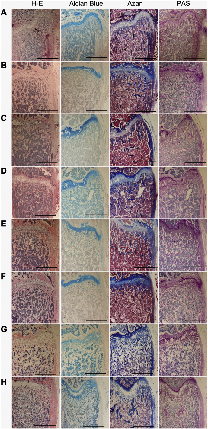
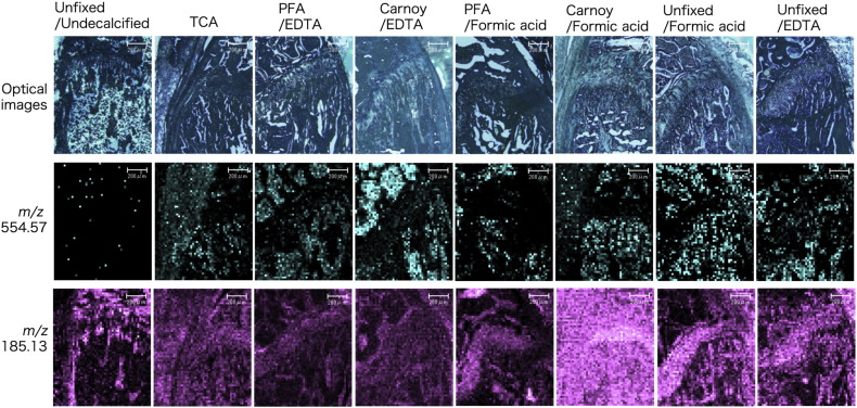
Matrix-assisted laser desorption/ionization-imaging mass spectrometry (MALDI-IMS) is an advanced method used globally to analyze the distribution of biomolecules on tissue cryosections without any probes. In bones, however, hydroxyapatite crystals make it difficult to determine the distribution of biomolecules using MALDI-IMS. Additionally, there is limited information regarding the use of this method to analyze bone tissues. To determine whether MALDI-IMS analysis of bone tissues can facilitate comprehensive mapping of biomolecules in mouse bone, we first dissected femurs and tibiae from 8-week-old male mice and characterized the quality of multiple fixation and decalcification methods for preparation of the samples. Cryosections were mounted on indium tin oxide-coated glass slides, dried, and then a matrix solution was sprayed on the tissue surface. Images were acquired using an iMScope at a mass-to-charge range of 100-1000. Hematoxylin-eosin, Alcian blue, Azan, and periodic acid-Schiff staining of adjacent sections was used to evaluate histological and histochemical features. Among the various fixation and decalcification conditions, sections from trichloroacetic acid-treated samples were most suitable to examine both histology and comprehensive MS images. However, histotypic MS signals were detected in all sections. In addition to the MS images, phosphocholine was identified as a candidate metabolite. These results indicate successful detection of biomolecules in bone using MALDI-IMS. Although analytical procedures and compositional adjustment regarding the performance of the device still require further development, IMS appears to be a powerful tool to determine the distribution of biomolecules in bone tissues.
基质辅助激光解吸/电离成像质谱(MALDI-IMS)是一种先进的方法,在全球范围内用于分析生物分子在组织冰冻切片上的分布,无需任何探针。然而,在骨骼中,羟基磷灰石晶体使得使用MALDI-IMS确定生物分子的分布变得困难。此外,关于使用该方法分析骨组织的信息有限。为了确定对骨组织进行MALDI-IMS分析是否有助于全面绘制小鼠骨骼中的生物分子图谱,我们首先从8周龄雄性小鼠身上解剖出股骨和胫骨,并对多种用于样本制备的固定和脱钙方法的质量进行了表征。将冰冻切片安装在涂有氧化铟锡的载玻片上,干燥,然后将基质溶液喷在组织表面。使用iMScope在质荷比范围为100-1000的条件下采集图像。对相邻切片进行苏木精-伊红、阿尔辛蓝、阿赞和过碘酸-希夫染色,以评估组织学和组织化学特征。在各种固定和脱钙条件中,三氯乙酸处理样本的切片最适合同时检查组织学和全面的质谱图像。然而,在所有切片中都检测到了组织型质谱信号。除了质谱图像外,磷酸胆碱被鉴定为一种候选代谢物。这些结果表明使用MALDI-IMS成功检测到了骨骼中的生物分子。尽管关于该设备性能的分析程序和成分调整仍需要进一步发展,但IMS似乎是确定骨组织中生物分子分布的有力工具。