Institute of Structural and Molecular Biology, Birkbeck College, Malet Street, London WC1E 7HX, England.
Acta Crystallogr D Struct Biol. 2017 Jun 1;73(Pt 6):509-521. doi: 10.1107/S2059798317007446. Epub 2017 May 31.
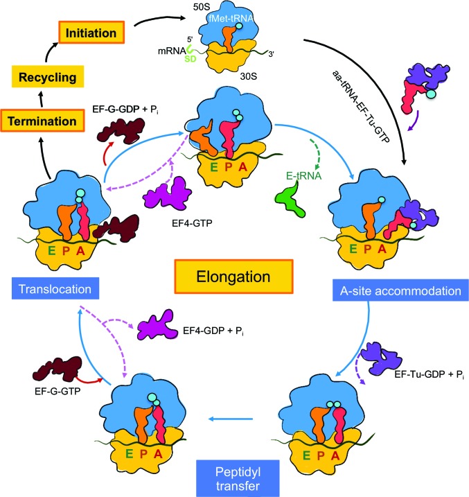
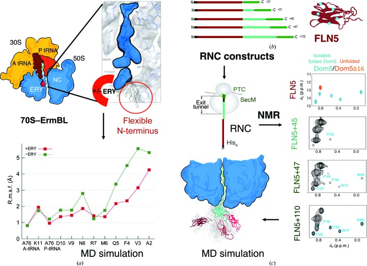
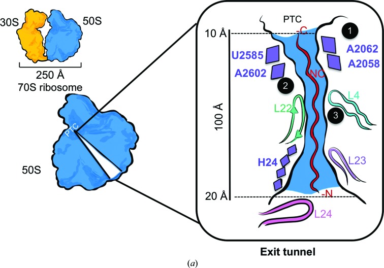
Protein folding, a process that underpins cellular activity, begins co-translationally on the ribosome. During translation, a newly synthesized polypeptide chain enters the ribosomal exit tunnel and actively interacts with the ribosome elements - the r-proteins and rRNA that line the tunnel - prior to emerging into the cellular milieu. While understanding of the structure and function of the ribosome has advanced significantly, little is known about the process of folding of the emerging nascent chain (NC). Advances in cryo-electron microscopy are enabling visualization of NCs within the exit tunnel, allowing early glimpses of the interplay between the NC and the ribosome. Once it has emerged from the exit tunnel into the cytosol, the NC (still attached to its parent ribosome) can acquire a range of conformations, which can be characterized by NMR spectroscopy. Using experimental restraints within molecular-dynamics simulations, the ensemble of NC structures can be described. In order to delineate the process of co-translational protein folding, a hybrid structural biology approach is foreseeable, potentially offering a complete atomic description of protein folding as it occurs on the ribosome.
蛋白质折叠是细胞活动的基础,它在核糖体上发生共翻译折叠。在翻译过程中,新合成的多肽链进入核糖体出口隧道,并在进入细胞环境之前与核糖体元件(排列在隧道中的 r 蛋白和 rRNA)积极相互作用。虽然对核糖体的结构和功能有了显著的了解,但对于新生链(NC)的折叠过程知之甚少。低温电子显微镜的进步使人们能够在出口隧道内可视化 NC,从而使人们能够早期了解 NC 与核糖体之间的相互作用。一旦从出口隧道进入细胞质,NC(仍然附着在其亲本核糖体上)可以获得一系列构象,可以通过 NMR 光谱进行表征。通过分子动力学模拟中的实验约束,可以描述 NC 结构的集合。为了描绘共翻译蛋白质折叠的过程,一种混合结构生物学方法是可以预见的,它可能提供蛋白质折叠过程的完整原子描述,就像它在核糖体上发生的那样。