• 文献检索
  • 文档翻译
  • 深度研究
  • 学术资讯
  • Suppr Zotero 插件Zotero 插件
  • 邀请有礼
  • 套餐&价格
  • 历史记录
应用&插件
Suppr Zotero 插件Zotero 插件浏览器插件Mac 客户端Windows 客户端微信小程序
定价
高级版会员购买积分包购买API积分包
服务
文献检索文档翻译深度研究API 文档MCP 服务
关于我们
关于 Suppr公司介绍联系我们用户协议隐私条款
关注我们

Suppr 超能文献

核心技术专利:CN118964589B侵权必究
粤ICP备2023148730 号-1Suppr @ 2026

文献检索

告别复杂PubMed语法,用中文像聊天一样搜索,搜遍4000万医学文献。AI智能推荐,让科研检索更轻松。

立即免费搜索

文件翻译

保留排版,准确专业,支持PDF/Word/PPT等文件格式,支持 12+语言互译。

免费翻译文档

深度研究

AI帮你快速写综述,25分钟生成高质量综述,智能提取关键信息,辅助科研写作。

立即免费体验

蝶腭孔快速定位的标志:影像学形态测量分析

Landmarks for rapid localization of the sphenopalatine foramen: A radiographic morphometric analysis.

作者信息

Maxwell Anne K, Barham Henry P, Getz Anne E, Kingdom Todd T, Ramakrishnan Vijay R

出版信息

Allergy Rhinol (Providence). 2017 Jun 1;8(2):63-66. doi: 10.2500/ar.2017.8.0196.

DOI:10.2500/ar.2017.8.0196
PMID:28583229
原文链接:https://pmc.ncbi.nlm.nih.gov/articles/PMC5468758/
Abstract

BACKGROUND

Transnasal endoscopic sphenopalatine artery ligation is becoming the procedure of choice for surgical management of intractable posterior epistaxis. Landmarks for localization of the sphenopalatine foramen can assist in rapid surgical exposure of the sphenopalatine artery.

OBJECTIVE

This study examined distances from easily identified endoscopic surgical landmarks to the sphenopalatine foramen.

METHODS

By using computed tomography of the sinus to study radiologic anatomy in 50 adults, distances were measured between five simple endoscopic landmarks and the sphenopalatine foramen. The two-tailed t-test was used for statistical analysis.

RESULTS

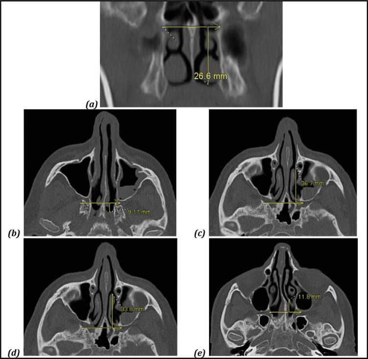
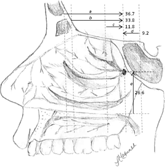
Right- and left-sided measurements were similar. The mean (standard deviation [SD]) anteroposterior distances to the sphenopalatine foramen were the following: from the maxillary line (36.7 ± 5.5 mm), anterior head of the middle turbinate (33.8 ± 6.7 mm), basal lamella (11.8 ± 1.9 mm), and choanal arch (-9.2 ± 1.4 mm). The mean (SD) distance in the vertical dimension from the nasal floor was 26.6 ± 2.6 mm. Female patients had statistically shorter distances to the sphenopalatine foramen from the maxillary line, anterior head of the middle turbinate, choanal arch, and nasal floor.

CONCLUSION

Reliable endoscopic landmarks exist in relation to consistent anatomic structures and can be used to help quickly estimate the location of the sphenopalatine foramen at the onset of the procedure.

摘要

背景

经鼻内镜蝶腭动脉结扎术正成为难治性后鼻孔出血手术治疗的首选方法。蝶腭孔的定位标志有助于快速手术暴露蝶腭动脉。

目的

本研究检测了从易于识别的内镜手术标志到蝶腭孔的距离。

方法

通过使用鼻窦计算机断层扫描研究50名成年人的放射解剖结构,测量了五个简单内镜标志与蝶腭孔之间的距离。采用双侧t检验进行统计分析。

结果

左右侧测量结果相似。到蝶腭孔的平均(标准差[SD])前后距离如下:从上颌线(36.7±5.5mm)、中鼻甲前端(33.8±6.7mm)、基板(11.8±1.9mm)和鼻后孔弓(-9.2±1.4mm)。从鼻底起垂直方向的平均(SD)距离为26.6±2.6mm。女性患者从上颌线、中鼻甲前端、鼻后孔弓和鼻底到蝶腭孔的距离在统计学上较短。

结论

与一致的解剖结构相关存在可靠的内镜标志,可用于在手术开始时帮助快速估计蝶腭孔的位置。

https://cdn.ncbi.nlm.nih.gov/pmc/blobs/1fec/5468758/fe8a3d97a172/arh0011701960002.jpg
https://cdn.ncbi.nlm.nih.gov/pmc/blobs/1fec/5468758/dbae0ae1f7d3/arh0011701960001.jpg
https://cdn.ncbi.nlm.nih.gov/pmc/blobs/1fec/5468758/fe8a3d97a172/arh0011701960002.jpg
https://cdn.ncbi.nlm.nih.gov/pmc/blobs/1fec/5468758/dbae0ae1f7d3/arh0011701960001.jpg
https://cdn.ncbi.nlm.nih.gov/pmc/blobs/1fec/5468758/fe8a3d97a172/arh0011701960002.jpg

相似文献

1
Landmarks for rapid localization of the sphenopalatine foramen: A radiographic morphometric analysis.蝶腭孔快速定位的标志:影像学形态测量分析
Allergy Rhinol (Providence). 2017 Jun 1;8(2):63-66. doi: 10.2500/ar.2017.8.0196.
2
Sphenopalatine Foramen Computed Tomography Landmarks.蝶腭孔的计算机断层扫描标志
J Craniofac Surg. 2020 Jan/Feb;31(1):210-213. doi: 10.1097/SCS.0000000000005857.
3
Survey Anatomical Features and Related Factors of the Sphenopalatine Foramen on Computed Tomography.计算机断层扫描下蝶腭孔的解剖学特征及相关因素研究
Indian J Otolaryngol Head Neck Surg. 2022 Oct;74(Suppl 2):1374-1378. doi: 10.1007/s12070-021-02494-z. Epub 2021 Mar 22.
4
Sphenopalatine foramen: endoscopic approach with bony landmarks.蝶腭孔:带骨性标志的内镜入路
J Laryngol Otol. 2015 Jul;129 Suppl 3:S47-52. doi: 10.1017/S0022215115000766. Epub 2015 Mar 30.
5
Endoscopic study for the pterygopalatine fossa anatomy: via the middle nasal meatus-sphenopalatine foramen approach.翼腭窝解剖结构的内镜研究:经中鼻道-蝶腭孔入路
J Craniofac Surg. 2009 May;20(3):944-7. doi: 10.1097/SCS.0b013e3181a2d9c8.
6
[Anatomic variations and references of the sphenopalatine foramen in cadaveric specimens: a Mexican study].[尸体标本中蝶腭孔的解剖变异及参考值:一项墨西哥的研究]
Cir Cir. 2014 Jul-Aug;82(4):367-71.
7
Anatomy of the sphenopalatine artery and its implications for transnasal neurosurgery.蝶腭动脉解剖及其对经鼻神经外科的意义。
Rhinology. 2018 Mar 1;56(1):82-88. doi: 10.4193/Rhin17.181.
8
Morphometric analysis of sphenopalatine foramen on computed tomography images with clinical significance.蝶腭孔 CT 影像形态学分析及其临床意义
Surg Radiol Anat. 2022 Dec;44(12):1521-1529. doi: 10.1007/s00276-022-03044-0. Epub 2022 Nov 17.
9
The role of the crista ethmoidalis in endoscopic sphenopalatine artery ligation.筛骨嵴在鼻内镜下蝶腭动脉结扎术中的作用
Am J Rhinol. 1999 Mar-Apr;13(2):81-6. doi: 10.2500/105065899782106814.
10
Mapping surgical coordinates of the sphenopalatine foramen: surgical navigation study.蝶腭孔手术坐标的定位:手术导航研究
J Laryngol Otol. 2009 Jul;123(7):742-5. doi: 10.1017/S0022215109004344. Epub 2009 Jan 8.

引用本文的文献

1
A Radiographic Morphometric Analysis for the Preoperative Localization of the Sphenopalatine Artery.蝶腭动脉术前定位的影像学形态计量分析
Indian J Otolaryngol Head Neck Surg. 2025 Mar;77(3):1424-1428. doi: 10.1007/s12070-025-05354-2. Epub 2025 Jan 20.
2
Survey Anatomical Features and Related Factors of the Sphenopalatine Foramen on Computed Tomography.计算机断层扫描下蝶腭孔的解剖学特征及相关因素研究
Indian J Otolaryngol Head Neck Surg. 2022 Oct;74(Suppl 2):1374-1378. doi: 10.1007/s12070-021-02494-z. Epub 2021 Mar 22.
3
Morphological Description and Clinical Implication of Sphenopalatine Foramen for Accurate Transnasal Sphenopalatine Ganglion Block: An Anatomical Study.

本文引用的文献

1
European position paper on the anatomical terminology of the internal nose and paranasal sinuses.欧洲关于内鼻和鼻窦解剖学术语的立场文件。
Rhinol Suppl. 2014 Mar;24:1-34.
2
Management of intractable spontaneous epistaxis.难治性自发性鼻出血的处理。
Am J Rhinol Allergy. 2012 Jan-Feb;26(1):55-60. doi: 10.2500/ajra.2012.26.3696.
3
Endoscopic ligation of the sphenopalatine artery and the maxillary artery for the treatment of intractable posterior epistaxis.内镜下结扎蝶腭动脉和上颌动脉治疗难治性鼻出血
用于精确经鼻蝶腭神经节阻滞的蝶腭孔的形态学描述及临床意义:一项解剖学研究
Medeni Med J. 2019;34(3):239-243. doi: 10.5222/MMJ.2019.20586. Epub 2019 Sep 27.
Am J Rhinol Allergy. 2009 Mar-Apr;23(2):197-9. doi: 10.2500/ajra.2009.23.3294.
4
Variations in paranasal sinus anatomy: implications for the pathophysiology of chronic rhinosinusitis and safety of endoscopic sinus surgery.鼻窦解剖结构的变异:对慢性鼻-鼻窦炎病理生理学及鼻内镜鼻窦手术安全性的影响
J Otolaryngol Head Neck Surg. 2009 Feb;38(1):32-7.
5
Mapping surgical coordinates of the sphenopalatine foramen: surgical navigation study.蝶腭孔手术坐标的定位:手术导航研究
J Laryngol Otol. 2009 Jul;123(7):742-5. doi: 10.1017/S0022215109004344. Epub 2009 Jan 8.
6
Changes in nasal cavity dimensions in children and adults by gender and age.儿童和成人鼻腔尺寸按性别和年龄的变化。
Laryngoscope. 2007 Aug;117(8):1429-33. doi: 10.1097/MLG.0b013e318064e837.
7
Update on epistaxis.鼻出血的最新情况。
Curr Opin Otolaryngol Head Neck Surg. 2007 Jun;15(3):180-3. doi: 10.1097/MOO.0b013e32814b06ed.
8
The relationship between anatomic variations of paranasal sinuses and chronic sinusitis in children.儿童鼻窦解剖变异与慢性鼻窦炎之间的关系。
Acta Otolaryngol. 2006 Oct;126(10):1067-72. doi: 10.1080/00016480600606681.
9
Posterior epistaxis: systematic review on the effectiveness of surgical therapies.后鼻孔出血:手术治疗效果的系统评价
Rhinology. 2005 Dec;43(4):300-4.
10
Endoscopic sphenopalatine artery ligation--when, why and how to do it. An on-line video tutorial.内镜下蝶腭动脉结扎术——时机、原因及操作方法。在线视频教程。
Clin Otolaryngol. 2005 Dec;30(6):539-43. doi: 10.1111/j.1749-4486.2005.01108.x.