Center for Alzheimer's and Neurodegenerative Diseases, Peter O'Donnell Jr. Brain Institute, University of Texas Southwestern Medical Center, Dallas, TX, USA.
Graduate Program in Neuroscience, Washington University in St. Louis, St. Louis, MO, USA.
Acta Neuropathol Commun. 2017 Jun 7;5(1):41. doi: 10.1186/s40478-017-0442-8.
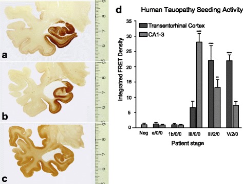
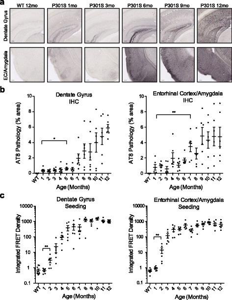
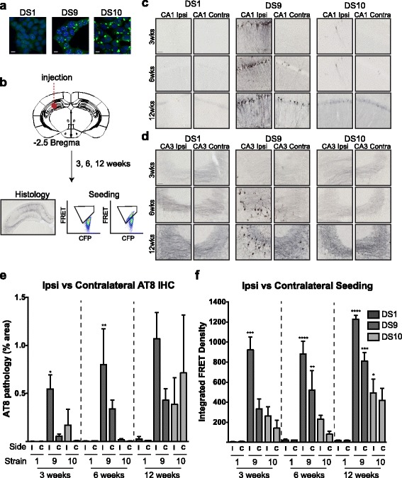
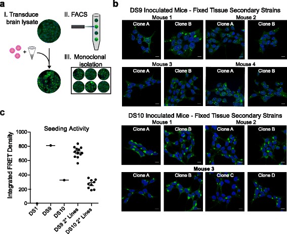
Tauopathies such as Alzheimer's disease (AD) feature progressive intraneuronal deposition of aggregated tau protein. The cause is unknown, but in experimental systems trans-cellular propagation of tau pathology resembles prion pathogenesis. Tau aggregate inoculation into mice produces transmissible pathology, and tau forms distinct strains, i.e. conformers that faithfully replicate and create predictable patterns of pathology in vivo. The prion model predicts that tau seed formation will anticipate neurofibrillary tau pathology. To test this idea requires simultaneous assessment of seed titer and immunohistochemistry (IHC) of brain tissue, but it is unknown whether tau seed titer can be determined in formaldehyde-fixed tissue. We have previously created a cellular biosensor system that uses flow cytometry to quantify induced tau aggregation and thus determine seed titer. In unfixed tissue from PS19 tauopathy mice that express 1 N,4R tau (P301S), we have measured tau seeding activity that precedes the first observable histopathology by many months. Additionally, in fresh frozen tissue from human AD subjects at early to mid-neurofibrillary tangle stages (NFT I-IV), we have observed tau seeding activity in cortical regions predicted to lack neurofibrillary pathology. However, we could not directly compare the same regions by IHC and seeding activity in either case. We now describe a protocol to extract and measure tau seeding activity from small volumes (.04 mm) of formaldehyde-fixed tissue immediately adjacent to that used for IHC. We validated this method with the PS19 transgenic mouse model, and easily observed seeding well before the development of phospho-tau pathology. We also accurately isolated two tau strains, DS9 and DS10, from fixed brain tissues in mice. Finally, we have observed robust seeding activity in fixed AD brain, but not controls. The successful coupling of classical IHC with seeding and strain detection should enable detailed study of banked brain tissue in AD and other tauopathies.
tau 病,如阿尔茨海默病 (AD),其特征是神经细胞内 tau 蛋白的聚集体逐渐沉积。其病因未知,但在实验系统中,tau 病理学的跨细胞传播类似于朊病毒发病机制。tau 聚集物接种到小鼠中会产生可传播的病理学,并且 tau 形成独特的株系,即能够忠实复制并在体内产生可预测的病理学模式的构象。朊病毒模型预测 tau 种子的形成将先于神经纤维缠结 tau 病理学。要验证这一想法,需要同时评估种子滴度和脑组织的免疫组织化学 (IHC),但尚不清楚是否可以在甲醛固定的组织中确定 tau 种子滴度。我们之前创建了一种细胞生物传感器系统,该系统使用流式细胞术来量化诱导的 tau 聚集,从而确定种子滴度。在表达 1N,4R tau (P301S) 的 PS19 tau 病小鼠的未固定组织中,我们已经测量了 tau 种子活性,该活性在数月前首次观察到组织病理学之前就已经出现。此外,在处于早期到中期神经纤维缠结 (NFT I-IV) 阶段的人类 AD 患者的新鲜冷冻组织中,我们在皮质区域观察到了 tau 种子活性,这些区域预计缺乏神经纤维病理学。然而,在任何情况下,我们都无法通过 IHC 和种子活性直接比较相同的区域。现在,我们描述了一种从紧邻用于 IHC 的福尔马林固定组织中提取和测量 tau 种子活性的小体积(0.04mm)的方案。我们使用 PS19 转基因小鼠模型验证了该方法,并且很容易在磷酸化 tau 病理学发展之前观察到种子。我们还从固定的小鼠脑组织中准确地分离出了两种 tau 株系 DS9 和 DS10。最后,我们在固定的 AD 脑中观察到了强烈的种子活性,但在对照中没有观察到。经典 IHC 与种子和株系检测的成功结合,应该能够对 AD 和其他 tau 病的储存脑组织进行详细研究。