• 文献检索
  • 文档翻译
  • 深度研究
  • 学术资讯
  • Suppr Zotero 插件Zotero 插件
  • 邀请有礼
  • 套餐&价格
  • 历史记录
应用&插件
Suppr Zotero 插件Zotero 插件浏览器插件Mac 客户端Windows 客户端微信小程序
定价
高级版会员购买积分包购买API积分包
服务
文献检索文档翻译深度研究API 文档MCP 服务
关于我们
关于 Suppr公司介绍联系我们用户协议隐私条款
关注我们

Suppr 超能文献

核心技术专利:CN118964589B侵权必究
粤ICP备2023148730 号-1Suppr @ 2026

文献检索

告别复杂PubMed语法,用中文像聊天一样搜索,搜遍4000万医学文献。AI智能推荐,让科研检索更轻松。

立即免费搜索

文件翻译

保留排版,准确专业,支持PDF/Word/PPT等文件格式,支持 12+语言互译。

免费翻译文档

深度研究

AI帮你快速写综述,25分钟生成高质量综述,智能提取关键信息,辅助科研写作。

立即免费体验

多房棘球绦虫(1863年,吕卡特;圆叶目:带科)卵膜的起源、分化及功能超微结构

Origin, differentiation and functional ultrastructure of egg envelopes in the cestode Echinococcus multilocularis Leuckart, 1863 (Cyclophyllidea: Taeniidae).

作者信息

Świderski Zdzisław, Miquel Jordi, Azzouz-Maache Samira, Pétavy Anne-Françoise

机构信息

Witold Stefański Institute of Parasitology, Polish Academy of Sciences, 51/55 Twarda Street, 00-818, Warszawa, Poland.

Secció de Parasitologia, Departament de Biologia, Sanitat i Medi Ambient, Facultat de Farmàcia i Ciències de l'Alimentació, Universitat de Barcelona, Av. Joan XXIII, sn, 08028, Barcelona, Spain.

出版信息

Parasitol Res. 2017 Jul;116(7):1963-1971. doi: 10.1007/s00436-017-5479-x. Epub 2017 Jun 7.

DOI:10.1007/s00436-017-5479-x
PMID:28593390
原文链接:https://pmc.ncbi.nlm.nih.gov/articles/PMC5486576/
Abstract

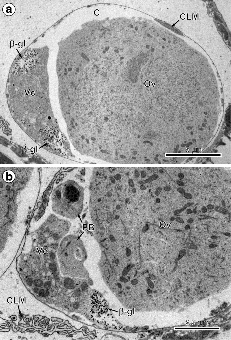
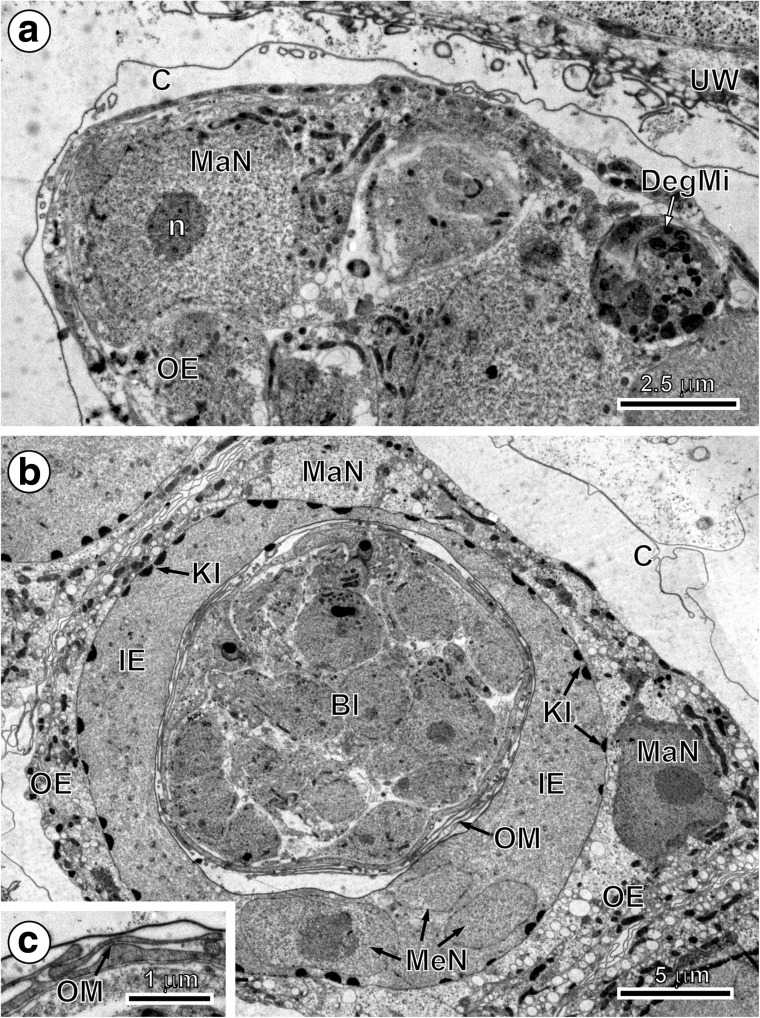
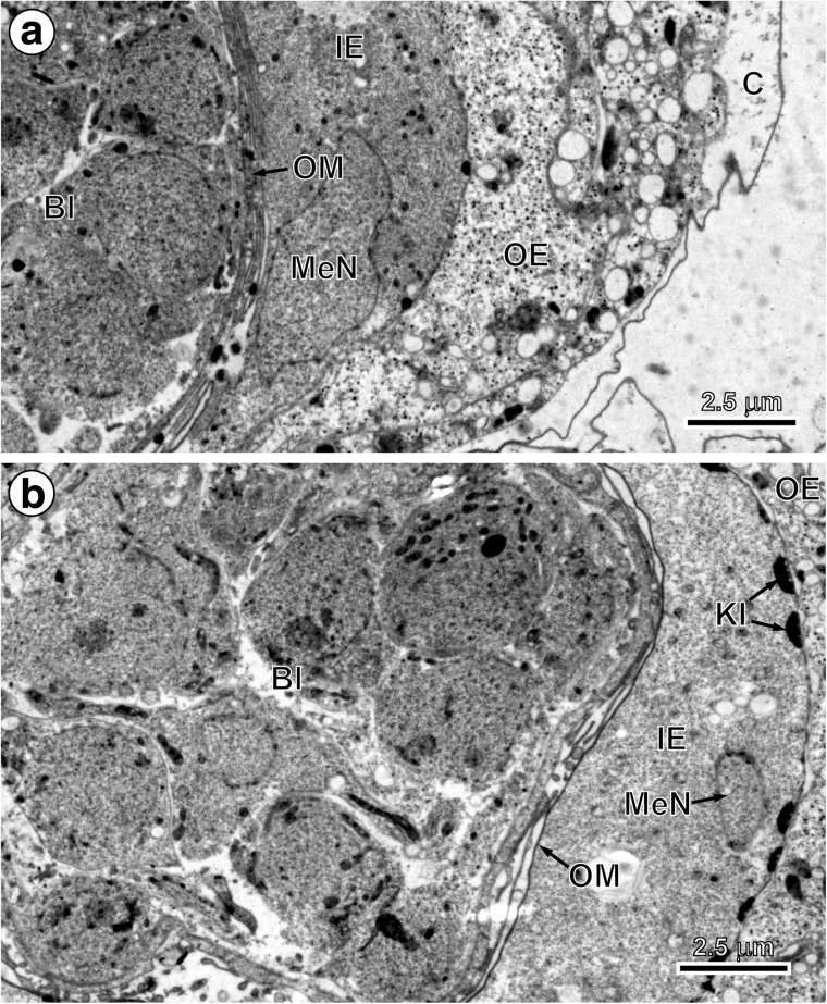
The origin, differentiation and functional ultrastructure of oncospheral or egg envelopes in Echinococcus multilocularis Leuckart, 1863 were studied by transmission electron microscopy (TEM) and cytochemistry. The purpose of our study is to describe the formation of the four primary embryonic envelopes, namely vitelline capsule, outer envelope, inner envelope and oncospheral membrane, and their transformation into the oncospheral or egg envelopes surrounding the mature hexacanth. This transformation takes place in the preoncospheral phase of embryonic development. The vitelline capsule and oncospheral membrane are thin membranes, while the outer and inner envelopes are thick cytoplasmic layers formed by two specific types of blastomeres: the outer envelope by cytoplasmic fusion of two macromeres and the inner envelope by cytoplasmic fusion of three mesomeres. Both outer and inner envelopes are therefore cellular in origin and syncytial in nature. During the advanced phase of embryonic development, the outer and inner envelopes undergo great modifications. The outer envelope remains as a metabolically active layer involved in the storage of glycogen and lipids for the final stages of egg development and survival. The inner envelope is the most important protective layer because of its thick layer of embryophoric blocks that assures oncospheral protection and survival. This embryophore is the principal layer of mature eggs, affording physical and physiological protection for the differentiated embryo or oncosphere, since the outer envelope is stripped from the egg before it is liberated. The embryophore is very thick and impermeable, consisting of polygonal blocks of an inert keratin-like protein held together by a cementing substance. The embryophore therefore assures extreme resistance of eggs, enabling them to withstand a wide range of environmental temperatures and physicochemical conditions.

摘要

利用透射电子显微镜(TEM)和细胞化学方法,对多房棘球绦虫(1863年,Leuckart)原头蚴或虫卵包膜的起源、分化及功能超微结构进行了研究。我们研究的目的是描述四种主要胚胎包膜的形成,即卵黄囊、外膜、内膜和原头蚴膜,以及它们如何转变为围绕成熟六钩蚴的原头蚴或虫卵包膜。这种转变发生在胚胎发育的原头蚴前期。卵黄囊和原头蚴膜是薄膜,而外膜和内膜是由两种特定类型的卵裂球形成的厚细胞质层:外膜由两个大卵裂球的细胞质融合形成,内膜由三个中卵裂球的细胞质融合形成。因此,外膜和内膜均起源于细胞,本质上为多核体。在胚胎发育的后期阶段,外膜和内膜发生了重大变化。外膜作为一层代谢活跃的层保留下来,参与为虫卵发育和存活的最后阶段储存糖原和脂质。内膜是最重要的保护层,因为它有一层厚厚的胚膜块,可确保原头蚴得到保护并存活。这种胚膜是成熟虫卵的主要层,为分化的胚胎或原头蚴提供物理和生理保护,因为外膜在虫卵释放前就从虫卵上剥离了。胚膜非常厚且不透水,由多边形的惰性角蛋白样蛋白质块通过一种胶结物质连接在一起组成。因此,胚膜确保了虫卵具有极强的抵抗力,使其能够承受广泛的环境温度和理化条件。

https://cdn.ncbi.nlm.nih.gov/pmc/blobs/5b4f/5486576/d2fb65836fa4/436_2017_5479_Fig6_HTML.jpg
https://cdn.ncbi.nlm.nih.gov/pmc/blobs/5b4f/5486576/3b25a0946e4c/436_2017_5479_Fig1_HTML.jpg
https://cdn.ncbi.nlm.nih.gov/pmc/blobs/5b4f/5486576/299fbcd6c8e1/436_2017_5479_Fig2_HTML.jpg
https://cdn.ncbi.nlm.nih.gov/pmc/blobs/5b4f/5486576/d3fa23ad3820/436_2017_5479_Fig3_HTML.jpg
https://cdn.ncbi.nlm.nih.gov/pmc/blobs/5b4f/5486576/4234818c3ec6/436_2017_5479_Fig4_HTML.jpg
https://cdn.ncbi.nlm.nih.gov/pmc/blobs/5b4f/5486576/b99bf6e262be/436_2017_5479_Fig5_HTML.jpg
https://cdn.ncbi.nlm.nih.gov/pmc/blobs/5b4f/5486576/d2fb65836fa4/436_2017_5479_Fig6_HTML.jpg

相似文献

1
Origin, differentiation and functional ultrastructure of egg envelopes in the cestode Echinococcus multilocularis Leuckart, 1863 (Cyclophyllidea: Taeniidae).多房棘球绦虫(1863年,吕卡特;圆叶目:带科)卵膜的起源、分化及功能超微结构
Parasitol Res. 2017 Jul;116(7):1963-1971. doi: 10.1007/s00436-017-5479-x. Epub 2017 Jun 7.
2
[Embryonic development of the cestode Mosgovoyia ctenoides (Anoplocephalidae)].[绦虫莫氏无钩绦虫(裸头科)的胚胎发育]
Wiad Parazytol. 2007;53(2):153-5.
3
Echinococcus multilocularis (Cestoda, Cyclophyllidea, Taeniidae): origin, differentiation and functional ultrastructure of the oncospheral tegument and hook region membrane.多房棘球绦虫(绦虫纲,圆叶目,带科):六钩蚴皮层和钩区膜的起源、分化及功能超微结构
Parasitol Res. 2018 Mar;117(3):783-791. doi: 10.1007/s00436-018-5752-7. Epub 2018 Jan 15.
4
Egg ultrastructure of the amabiliid cestode Tatria biremis Kowalewski, 1904 (Cyclophyllidea, Amabiliidae), with an emphasis on the oncospheral envelopes.1904年发现的美丽绦虫Tatria biremis Kowalewski(圆叶目,美丽绦虫科)的卵超微结构,重点研究六钩蚴膜。
Acta Parasitol. 2013 Sep;58(3):269-77. doi: 10.2478/s11686-013-0138-6. Epub 2013 Aug 29.
5
An ultrastructural study of embryonic envelope formation in the anoplocephalid cestode Mosgovoyia ctenoides (Railliet, 1890) Beveridge, 1978.对无钩绦虫类莫氏绦虫(Mosgovoyia ctenoides (Railliet, 1890) Beveridge, 1978)胚胎包膜形成的超微结构研究。
Parasitol Res. 2005 Mar;95(4):243-51. doi: 10.1007/s00436-004-1276-4. Epub 2005 Jan 28.
6
Echinococcus multilocularis (Cestoda, Cyclophyllidea, Taeniidae): oncospheral hook morphogenesis.多房棘球绦虫(绦虫纲,圆叶目,带科):六钩蚴钩的形态发生
Parasitol Res. 2016 Oct;115(10):3715-21. doi: 10.1007/s00436-016-5131-1. Epub 2016 May 20.
7
Differentiation and ultrastructure of oncospheral and uterine envelopes in the nematotaeniid cestode, Nematotaenia dispar (Goeze, 1782).线虫绦虫Nematotaenia dispar(戈泽,1782年)六钩蚴和子宫膜的分化与超微结构
Int J Parasitol. 1997 Sep;27(9):1065-74. doi: 10.1016/s0020-7519(97)00059-3.
8
Echinococcus multilocularis (Cestoda, Cyclophyllidea, Taeniidae): functional ultrastructure of the penetration glands and nerve cells within the oncosphere.多房棘球绦虫(绦虫纲,圆叶目,带科):六钩蚴内穿透腺和神经细胞的功能超微结构
Parasitol Res. 2018 Aug;117(8):2653-2663. doi: 10.1007/s00436-018-5957-9. Epub 2018 Jun 9.
9
[Embryonic development of 2 Phyllobothriidae (Cestoda: Tetraphyllidea)].双叶槽科(绦虫纲:四叶目)的胚胎发育
Ann Parasitol Hum Comp. 1976 May-Jun;51(3):309-27. doi: 10.1051/parasite/1976513309.
10
Transmission electron micrograph studies of developing oncospheral envelopes of Fimbriaria fasciolaris (Hymenolepididae).豆状带绦虫(膜壳科)发育中六钩蚴包膜的透射电子显微镜研究。
Parasitol Res. 1991;77(6):503-8. doi: 10.1007/BF00928418.

引用本文的文献

1
Suitability of red fox () and golden jackal () as hosts of based on egg production characteristics and literature data on the intestinal ecosystems of carnivores.基于产蛋特征及有关食肉动物肠道生态系统的文献数据,赤狐()和金豺()作为 宿主的适宜性。
Curr Res Parasitol Vector Borne Dis. 2024 Oct 28;6:100225. doi: 10.1016/j.crpvbd.2024.100225. eCollection 2024.
2
Important nutrient sources and carbohydrate metabolism patterns in the growth and development of spargana.棘头虫生长发育中的重要营养来源和碳水化合物代谢模式。
Parasit Vectors. 2024 Feb 16;17(1):68. doi: 10.1186/s13071-024-06148-1.

本文引用的文献

1
Echinococcus multilocularis (Cestoda, Cyclophyllidea, Taeniidae): oncospheral hook morphogenesis.多房棘球绦虫(绦虫纲,圆叶目,带科):六钩蚴钩的形态发生
Parasitol Res. 2016 Oct;115(10):3715-21. doi: 10.1007/s00436-016-5131-1. Epub 2016 May 20.
2
Functional ultrastructure of eggs and cellular organization of hexacanths of the cyclophyllidean cestode Thysanotaenia congolensis: a phylogenetic implication of obtained results.刚果带绦虫(Thysanotaenia congolensis)虫卵的功能超微结构及六钩蚴的细胞组织:所得结果的系统发育意义
Parasitology. 2016 Mar;143(3):320-33. doi: 10.1017/S0031182015001560. Epub 2015 Dec 22.
3
Ultrastructural reconstruction of Taenia ovis oncospheres from serial sections.
绵羊带绦虫原头蚴的连续切片超微结构重建。
Int J Parasitol. 2010 Oct;40(12):1419-31. doi: 10.1016/j.ijpara.2010.04.011. Epub 2010 May 15.
4
The ultrastructure of taeniid cestode oncospheres and localization of host-protective antigens.带科绦虫类原头蚴的超微结构和宿主保护性抗原的定位。
Parasitology. 2010 Mar;137(3):521-35. doi: 10.1017/S0031182009991260. Epub 2009 Oct 16.
5
A standardised terminology of the embryonic envelopes and associated developmental stages of tapeworms (Platyhelminthes: Cestoda).绦虫(扁形动物门:绦虫纲)胚胎膜及相关发育阶段的标准化术语。
Folia Parasitol (Praha). 2008 Mar;55(1):42-52. doi: 10.14411/fp.2008.006.
6
An ultrastructural study of embryonic envelope formation in the anoplocephalid cestode Mosgovoyia ctenoides (Railliet, 1890) Beveridge, 1978.对无钩绦虫类莫氏绦虫(Mosgovoyia ctenoides (Railliet, 1890) Beveridge, 1978)胚胎包膜形成的超微结构研究。
Parasitol Res. 2005 Mar;95(4):243-51. doi: 10.1007/s00436-004-1276-4. Epub 2005 Jan 28.
7
ULTRASTRUCTURE OF DEVELOPING TAENIID EMBRYOPHORES AND ASSOCIATED STRUCTURES.发育中的带绦虫胚膜及其相关结构的超微结构
Exp Parasitol. 1965 Apr;16:207-16. doi: 10.1016/0014-4894(65)90045-7.
8
The use of lead citrate at high pH as an electron-opaque stain in electron microscopy.在电子显微镜检查中,将高pH值的柠檬酸铅用作电子不透明染色剂。
J Cell Biol. 1963 Apr;17(1):208-12. doi: 10.1083/jcb.17.1.208.
9
Egg-shell formation in trematodes and cestodes.吸虫和绦虫的卵壳形成。
Exp Parasitol. 1959 Jun;8(3):286-323. doi: 10.1016/0014-4894(59)90027-x.
10
Hymenolepis nana: the fine structure of the embryonic envelopes.微小膜壳绦虫:胚胎膜的精细结构
Parasitology. 1981 Jun;82(Pt 3):429-43. doi: 10.1017/s0031182000066968.