Challapalli Amarnath, Carroll Laurence, Aboagye Eric O
Department of Clinical Oncology, Bristol Cancer Institute, Horfield Road, Bristol, United Kingdom.
Department of Surgery and Cancer, Imperial College, GN1, Commonwealth Building, Hammersmith Hospital, Du Cane Road, London, W120NN United Kingdom.
Clin Transl Imaging. 2017;5(3):225-253. doi: 10.1007/s40336-017-0231-1. Epub 2017 May 11.
Hypoxia is a condition of insufficient oxygen to support metabolism which occurs when the vascular supply is interrupted, or when a tumour outgrows its vascular supply. It is a negative prognostic factor due to its association with an aggressive tumour phenotype and therapeutic resistance. This review provides an overview of hypoxia imaging with Positron emission tomography (PET), with an emphasis on the biological relevance, mechanism of action, highlighting advantages, and limitations of the currently available hypoxia radiotracers.
A comprehensive PubMed literature search was performed, identifying articles relating to biological significance and measurement of hypoxia, MRI methods, and PET imaging of hypoxia in preclinical and clinical settings, up to December 2016.
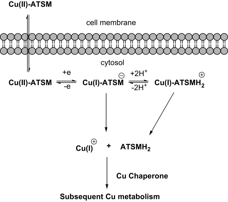
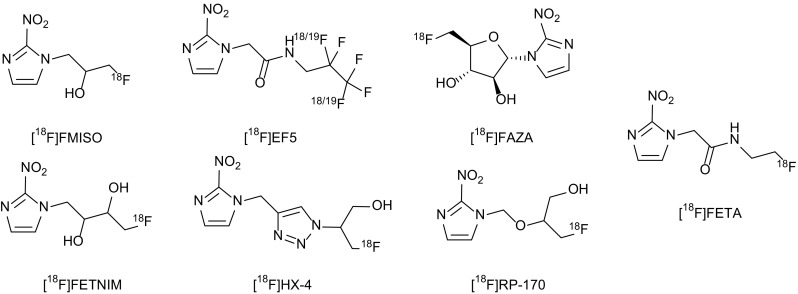
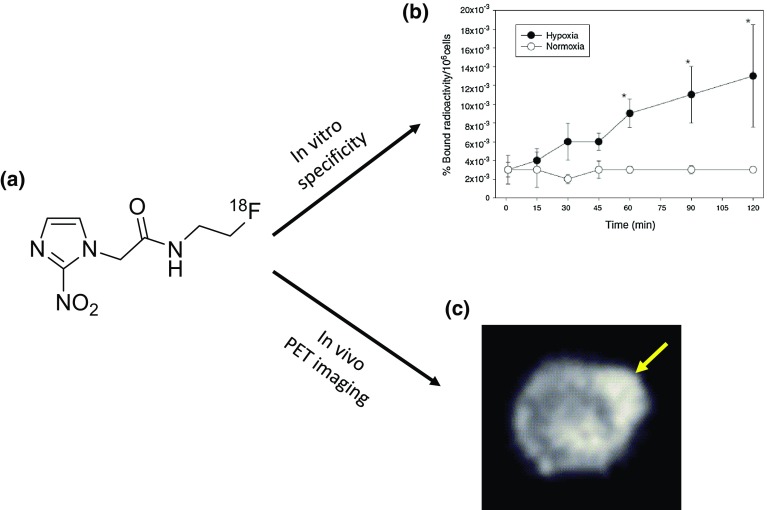
A variety of approaches have been explored over the years for detecting and monitoring changes in tumour hypoxia, including regional measurements with oxygen electrodes placed under CT guidance, MRI methods that measure either oxygenation or lactate production consequent to hypoxia, different nuclear medicine approaches that utilise imaging agents the accumulation of which is inversely related to oxygen tension, and optical methods. The advantages and disadvantages of these approaches are reviewed, along with individual strategies for validating different imaging methods. PET is the preferred method for imaging tumour hypoxia due to its high specificity and sensitivity to probe physiological processes in vivo, as well as the ability to provide information about intracellular oxygenation levels.
Even though hypoxia could have significant prognostic and predictive value in the clinic, the best method for hypoxia assessment has in our opinion not been realised.
缺氧是指氧气供应不足,无法维持新陈代谢的一种状态,当血管供应中断或肿瘤生长超过其血管供应时就会发生。由于它与侵袭性肿瘤表型及治疗耐药性相关,因此是一个负面的预后因素。本综述概述了正电子发射断层扫描(PET)用于缺氧成像的情况,重点介绍了当前可用的缺氧放射性示踪剂的生物学相关性、作用机制、突出优点及局限性。
进行了全面的PubMed文献检索,检索截至2016年12月的有关缺氧的生物学意义及测量、MRI方法以及临床前和临床环境中缺氧的PET成像的文章。
多年来已探索了多种检测和监测肿瘤缺氧变化的方法,包括在CT引导下用氧电极进行区域测量、测量缺氧导致的氧合或乳酸生成的MRI方法、利用成像剂且其积聚与氧张力呈负相关的不同核医学方法以及光学方法。对这些方法的优缺点进行了综述,并介绍了验证不同成像方法的具体策略。PET因其对体内探测生理过程具有高特异性和敏感性,以及能够提供有关细胞内氧合水平的信息,而成为成像肿瘤缺氧的首选方法。
尽管缺氧在临床上可能具有重要的预后和预测价值,但我们认为尚未找到评估缺氧的最佳方法。