Ishii Aya, Kimura Tokuhiro, Sadahiro Hirokazu, Kawano Hiroo, Takubo Keiyo, Suzuki Michiyasu, Ikeda Eiji
Department of Pathology, Yamaguchi University Graduate School of Medicine, Ube, Yamaguchi, Japan.
Department of Neurosurgery, Yamaguchi University Graduate School of Medicine, Ube, Yamaguchi, Japan.
PLoS One. 2016 Jan 22;11(1):e0147366. doi: 10.1371/journal.pone.0147366. eCollection 2016.
Characterization of the niches for stem-like tumor cells is important to understand and control the behavior of glioblastomas. Cell-cycle quiescence might be a common mechanism underlying the long-term maintenance of stem-cell function in normal and neoplastic stem cells, and our previous study demonstrated that quiescence induced by hypoxia-inducible factor (HIF)-1α is associated with a high long-term repopulation capacity of hematopoietic stem cells. Based on this, we examined human astrocytoma tissues for HIF-1α-regulated quiescent stem-like tumor cells as a candidate for long-term tumorigenic cells and characterized their niche histologically.
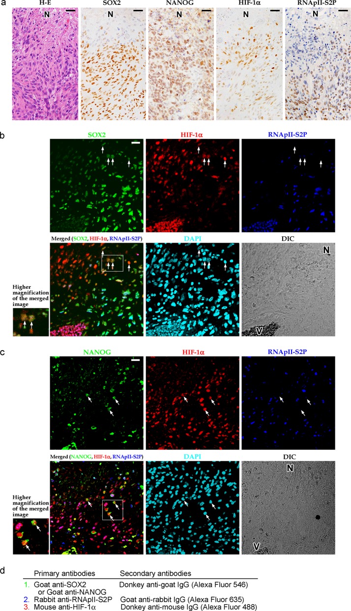
Multi-color immunohistochemistry was used to visualize HIF-1α-expressing (HIF-1α+) quiescent stem-like tumor cells and their niche in astrocytoma (WHO grade II-IV) tissues. This niche was modeled using spheroids of cultured glioblastoma cells and its contribution to tumorigenicity was evaluated by sphere formation assay.
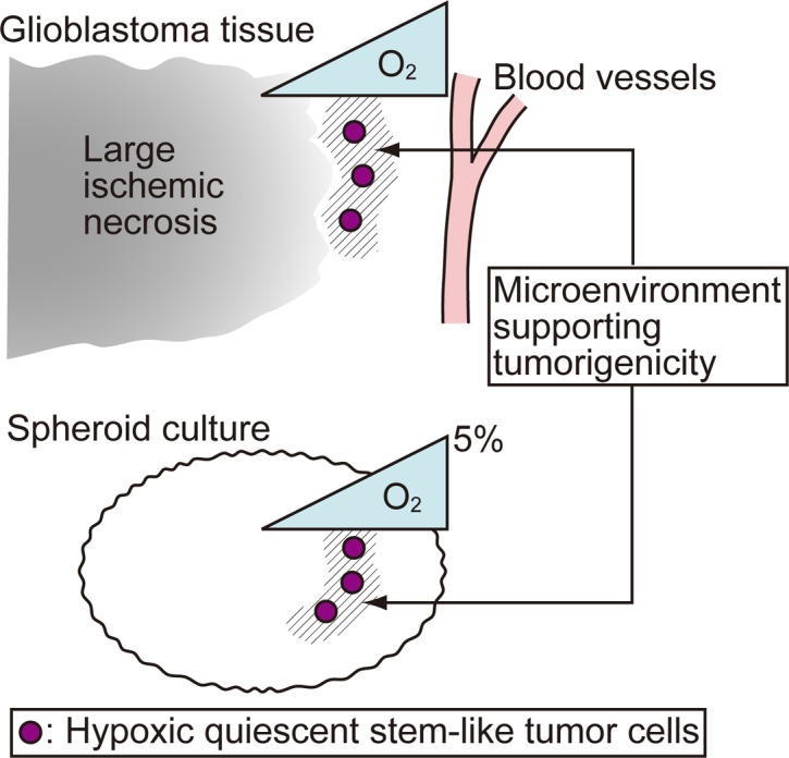
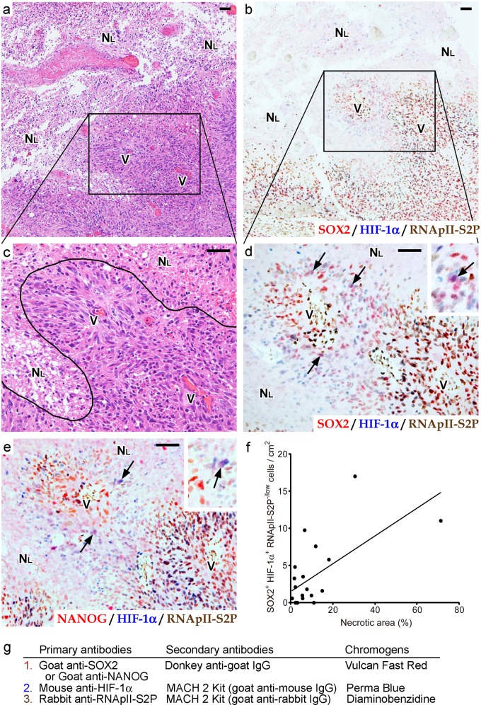
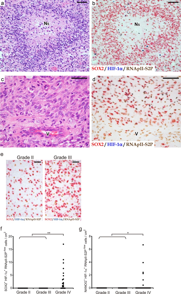
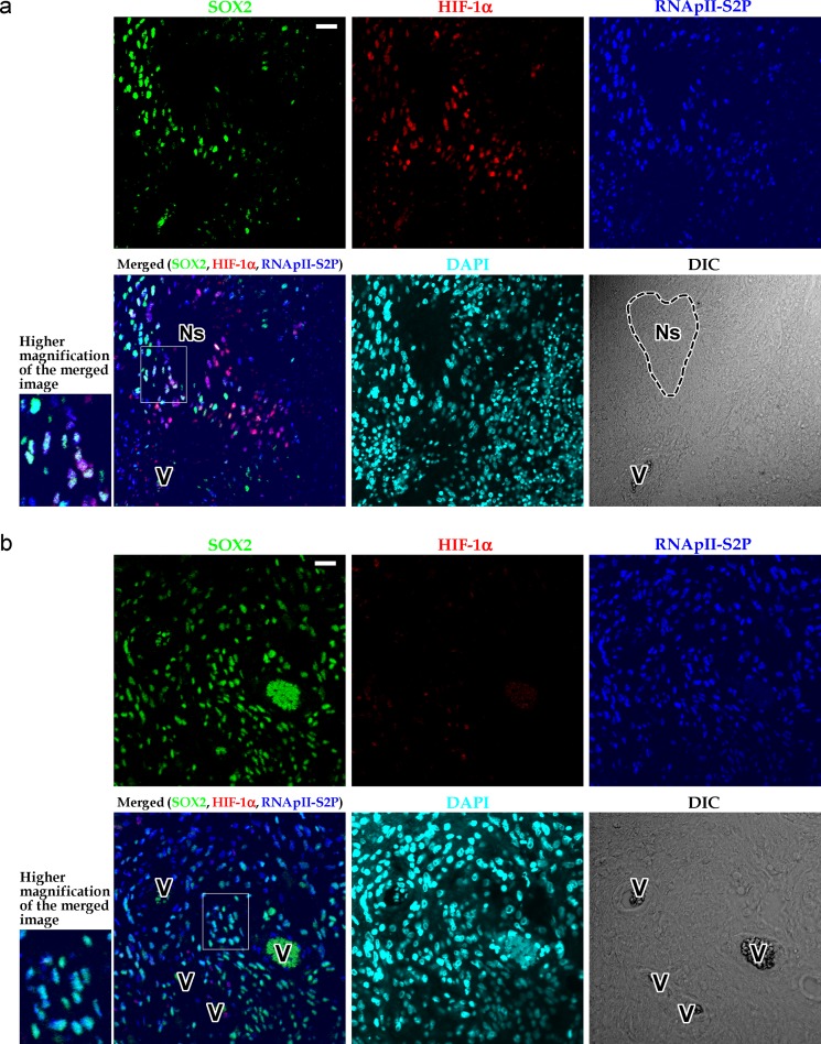
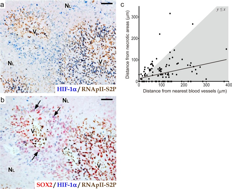
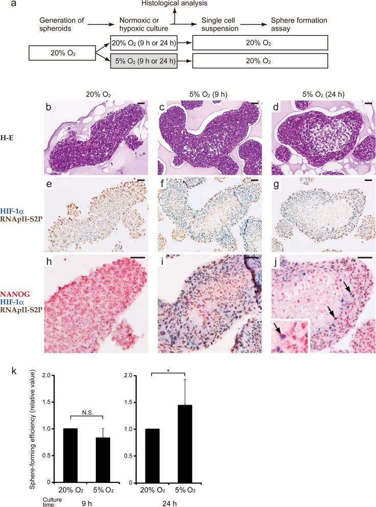
A small subpopulation of HIF-1α+ quiescent stem-like tumor cells was found in glioblastomas but not in lower-grade astrocytomas. These cells were concentrated in the zone between large ischemic necroses and blood vessels and were closer to the necrotic tissues than to the blood vessels, which suggested that a moderately hypoxic microenvironment is their niche. We successfully modeled this niche containing cells of HIF-1α+ quiescent stem-like phenotype by incubating glioblastoma cell spheroids under an appropriately hypoxic condition, and the emergence of HIF-1α+ quiescent stem-like cells was shown to be associated with an enhanced sphere-forming activity.
These data suggest that the "peri-necrotic niche" harboring HIF-1α+ quiescent stem-like cells confers a higher tumorigenic potential on glioblastoma cells and therefore may be a therapeutic target to control the behavior of glioblastomas.
对肿瘤干细胞龛进行特征描述对于理解和控制胶质母细胞瘤的行为很重要。细胞周期静止可能是正常干细胞和肿瘤干细胞长期维持干细胞功能的共同机制,并且我们之前的研究表明,缺氧诱导因子(HIF)-1α诱导的静止与造血干细胞的高长期再增殖能力相关。基于此,我们检测了人类星形细胞瘤组织中HIF-1α调节的静止肿瘤干细胞样细胞,将其作为长期致瘤细胞的候选者,并从组织学上对其龛进行了特征描述。
采用多色免疫组织化学法观察星形细胞瘤(世界卫生组织II-IV级)组织中表达HIF-1α(HIF-1α+)的静止肿瘤干细胞样细胞及其龛。使用培养的胶质母细胞瘤细胞球体对该龛进行建模,并通过球体形成试验评估其对致瘤性的贡献。
在胶质母细胞瘤中发现了一小部分HIF-1α+静止肿瘤干细胞样细胞,而在低级别星形细胞瘤中未发现。这些细胞集中在大的缺血性坏死灶和血管之间的区域,且更靠近坏死组织而非血管,这表明适度缺氧的微环境是它们的龛。通过在适当缺氧条件下培养胶质母细胞瘤细胞球体,我们成功地对包含HIF-1α+静止肿瘤干细胞样表型细胞的龛进行了建模,并且显示HIF-1α+静止肿瘤干细胞样细胞的出现与增强的球体形成活性相关。
这些数据表明,含有HIF-1α+静止肿瘤干细胞样细胞的 “坏死周龛” 赋予胶质母细胞瘤细胞更高的致瘤潜能,因此可能是控制胶质母细胞瘤行为的治疗靶点。