Hurtado-de-Mendoza David, Corona-Villalobos Celia P, Pozios Iraklis, Gonzales Jorge, Soleimanifard Yalda, Sivalokanathan Sanjay, Montoya-Cerrillo Diego, Vakrou Styliani, Kamel Ihab, Mormontoy-Laurel Wilfredo, Dolores-Cerna Ketty, Suarez Jacsel, Perez-Melo Sergio, Bluemke David A, Abraham Theodore P, Zimmerman Stefan L, Abraham M Roselle
Hypertrophic Cardiomyopathy Center of Excellence, Department of Medicine, Johns Hopkins University School of Medicine, 720 Rutland Ave, Ross 871, Baltimore, MD 21205, USA.
Cayetano Heredia University School of Medicine, 430 Honorio Delgado Ave, Lima, LIMA 31, Peru.
J Arrhythm. 2017 Jun;33(3):201-207. doi: 10.1016/j.joa.2016.10.005. Epub 2016 Nov 19.
Hypertrophic cardiomyopathy (HCM) is characterized by myocyte hypertrophy, disarray, fibrosis, and increased risk for ventricular arrhythmias. Increased QT dispersion has been reported in patients with HCM, but the underlying mechanisms have not been completely elucidated. In this study, we examined the relationship between diffuse interstitial fibrosis, replacement fibrosis, QTc dispersion and ventricular arrhythmias in patients with HCM. We hypothesized that fibrosis would slow impulse propagation and increase dispersion of ventricular repolarization, resulting in increased QTc dispersion on surface electrocardiogram (ECG) and ventricular arrhythmias.
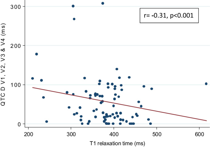
ECG and cardiac magnetic resonance (CMR) image analyses were performed retrospectively in 112 patients with a clinical diagnosis of HCM. Replacement fibrosis was assessed by measuring late gadolinium (Gd) enhancement (LGE), using a semi-automated threshold technique. Diffuse interstitial fibrosis was assessed by measuring T1 relaxation times after Gd administration, using the Look-Locker sequence. QTc dispersion was measured digitally in the septal/anterior (V1-V4), inferior (II, III, and aVF), and lateral (I, aVL, V5, and V6) lead groups on surface ECG.
All patients had evidence of asymmetric septal hypertrophy. LGE was evident in 70 (63%) patients; the median T1 relaxation time was 411±38 ms. An inverse correlation was observed between T1 relaxation time and QTc dispersion in leads V1-V4 (<0.001). Patients with HCM who developed sustained ventricular tachycardia had slightly higher probability of increased QTc dispersion in leads V1-V4 (odds ratio, 1.011 [1.004-1.0178, =0.003). We found no correlation between presence and percentage of LGE and QTc dispersion.
Diffuse interstitial fibrosis is associated with increased dispersion of ventricular repolarization in leads, reflecting electrical activity in the hypertrophied septum. Interstitial fibrosis combined with ion channel/gap junction remodeling in the septum could lead to inhomogeneity of ventricular refractoriness, resulting in increased QTc dispersion in leads V1-V4.
肥厚型心肌病(HCM)的特征是心肌细胞肥大、排列紊乱、纤维化以及室性心律失常风险增加。已有报道称HCM患者的QT离散度增加,但其潜在机制尚未完全阐明。在本研究中,我们探讨了HCM患者弥漫性间质纤维化、替代性纤维化、QTc离散度与室性心律失常之间的关系。我们假设纤维化会减慢冲动传导并增加心室复极离散度,导致体表心电图(ECG)上的QTc离散度增加和室性心律失常。
对112例临床诊断为HCM的患者进行回顾性ECG和心脏磁共振(CMR)图像分析。采用半自动阈值技术通过测量钆延迟增强(LGE)来评估替代性纤维化。使用Look-Locker序列通过测量钆给药后的T1弛豫时间来评估弥漫性间质纤维化。在体表ECG的间隔/前壁(V1-V4)、下壁(II、III和aVF)和侧壁(I、aVL、V5和V6)导联组中数字测量QTc离散度。
所有患者均有不对称性室间隔肥厚的证据。70例(63%)患者出现LGE;T1弛豫时间中位数为411±38 ms。在V1-V4导联中观察到T1弛豫时间与QTc离散度呈负相关(<0.001)。发生持续性室性心动过速的HCM患者在V-1V4导联中QTc离散度增加的可能性略高(优势比,1.011 [1.004-1.0178,P=0.003])。我们发现LGE的存在和百分比与QTc离散度之间无相关性。
弥漫性间质纤维化与导联中室性复极离散度增加相关,反映了肥厚室间隔中的电活动。间质纤维化与间隔中的离子通道/缝隙连接重塑相结合可能导致心室不应期的不均匀性,从而导致V1-V4导联中QTc离散度增加。