Barczuk-Falęcka Marzena, Bombiński Przemysław, Majkowska Zofia, Brzewski Michał, Warchoł Stanisław
Department of Pediatric Radiology, Medical University of Warsaw, Warsaw, Poland.
Department of Pediatric Surgery and Urology, Medical University of Warsaw, Warsaw, Poland.
Pol J Radiol. 2017 May 19;82:275-278. doi: 10.12659/PJR.899995. eCollection 2017.
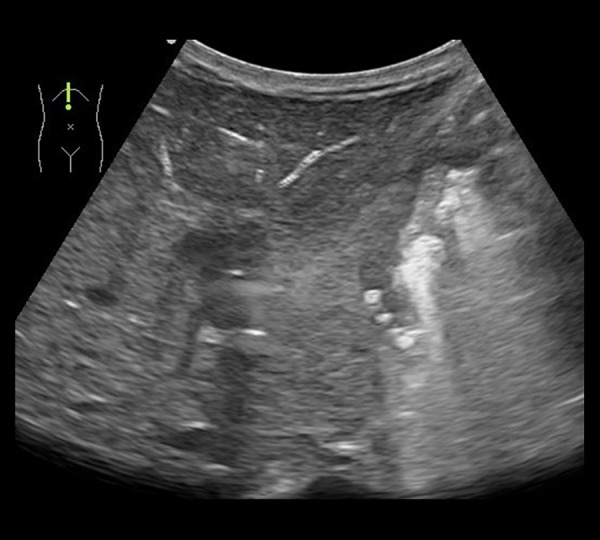
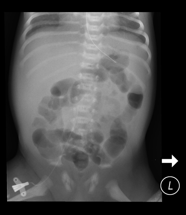
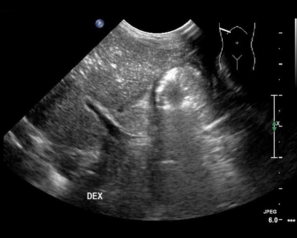
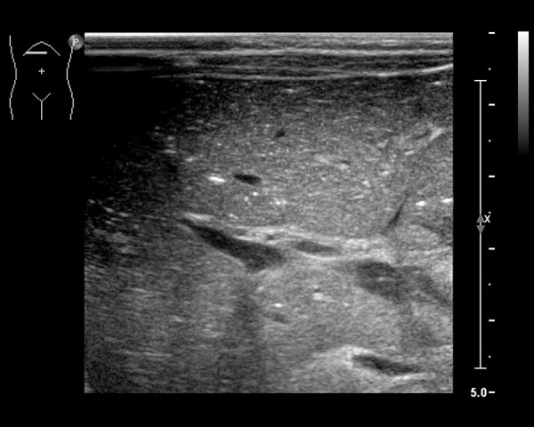
Hepatic portal venous gas (HPVG) is a rare imaging finding in children. It can be an important manifestation of severe diseases such as necrotizing enterocolitis (NEC) in neonates or bowel wall rupture in older children. However, there are many other diseases presenting with HPVG that do not necessarily require a surgical intervention.
In the period between 2011-2015, there were 12 cases of HPVG in children aged up to 24 months in our hospital. We did not include children with NEC. We retrospectively analyzed clinical data and US examinations as regards the suspected causes and final diagnoses. Only 1 patient with HPVG required an immediate surgical intervention. This was - a 4-month-old girl 32 days after a repair of a congenital diaphragmatic hernia, with ultrasound signs of acute bowel wall necrosis. During surgery a bowel strangulation was revealed. Other causes included: - 4 patients with bowel inflammation (including complications of neoplastic diseases such as leukemia and Hodgkins'disease); - 3 patients with food allergy; - 1 patient with acute gastroenteritis; - 1 patient with hepatic injury because of a suspected metabolic disease; - 1 incidental finding revealed before closing a ventricular septum defect; - 1 patient during follow-up performed 2 weeks after a reconstruction of bowel continuity.
HPVG is not always a sign of a life-threatening condition and it should not be by itself an indication for surgical treatment. HPVG requires a close monitoring of the clinical status, which is crucial for further management. In patients in non-severe clinical condition, we propose to perform a follow-up ultrasound imaging within 1-2 days, and not to extend diagnostic procedures, especially in case of ultrasound picture normalization. An abdominal ultrasound examination appears to be the method of choice for the identification of gas in the hepatic portal system in children.
肝门静脉积气(HPVG)在儿童中是一种罕见的影像学表现。它可能是新生儿坏死性小肠结肠炎(NEC)或大龄儿童肠壁破裂等严重疾病的重要表现。然而,还有许多其他疾病也可出现HPVG,并不一定需要手术干预。
2011年至2015年期间,我院有12例24个月以下儿童出现HPVG。我们未纳入患有NEC的儿童。我们回顾性分析了关于疑似病因和最终诊断的临床资料及超声检查结果。只有1例HPVG患儿需要立即进行手术干预。该患儿为一名4个月大的女孩,在先天性膈疝修补术后32天,超声显示有急性肠壁坏死迹象。手术中发现肠绞窄。其他病因包括:4例肠炎患者(包括白血病和霍奇金病等肿瘤性疾病的并发症);3例食物过敏患者;1例急性胃肠炎患者;1例因疑似代谢性疾病导致肝损伤的患者;1例在室间隔缺损修补术关闭前偶然发现;1例在肠连续性重建术后2周随访期间发现。
HPVG并不总是危及生命状况的征象,其本身不应作为手术治疗的指征。HPVG需要密切监测临床状况,这对进一步的处理至关重要。对于临床状况不严重的患者,我们建议在1 - 2天内进行超声随访检查,且不要扩大诊断程序,尤其是在超声图像正常化的情况下。腹部超声检查似乎是识别儿童肝门静脉系统气体的首选方法。