Cavaignac Etienne, Savall Frederic, Chantalat Elodie, Faruch Marie, Reina Nicolas, Chiron Philippe, Telmon Norbert
Laboratoire AMIS, UMR 5288 CNRS, Université Paul Sabatier, 37 allée Jules Guesde, 31000, Toulouse, France.
Institut de l'appareil locomoteur, Hôpital Pierre-Paul Riquet, CHU Toulouse, France.
J Exp Orthop. 2017 Dec;4(1):21. doi: 10.1186/s40634-017-0095-3. Epub 2017 Jun 12.
Few studies have looked into age-related variations in femur shape. We hypothesized that three-dimensional (3D) geometric morphometric analysis of the distal femur would reveal age-related differences. The purpose of this study was to show that differences in distal femur shape related to age could be identified, visualized, and quantified using three-dimensional (3D) geometric morphometric analysis.
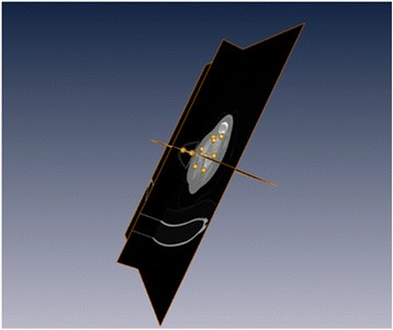
Geometric morphometric analysis was carried out on CT scans of the distal femur of 256 subjects living in the south of France. Ten landmarks were defined on 3D reconstructions of the distal femur. Both traditional metric and geometric morphometric analyses were carried out on these bone reconstructions. These analyses were used to identify trends in bone shape in various age-based subgroups (<40, 40-60, >60).
Only the average bone shape of the < 40-year subgroup was statistically different from that of the other two groups. When the population was divided into two subgroups using 40 years of age as a threshold, the subject's age was correctly assigned 80% of the time.
Age-related differences are present in this bone segment. This reliable, accurate method could be used for virtual autopsy and to perform diachronic and interethnic comparisons. Moreover, this study provides updated morphometric data for a modern population in the south of France.
Manufacturers of knee replacement implants will have to adapt their prosthesis models as the population evolves over time.
很少有研究探讨股骨形状与年龄相关的变化。我们假设对股骨远端进行三维(3D)几何形态计量分析将揭示与年龄相关的差异。本研究的目的是表明,使用三维(3D)几何形态计量分析可以识别、可视化和量化与年龄相关的股骨远端形状差异。
对居住在法国南部的256名受试者的股骨远端CT扫描进行几何形态计量分析。在股骨远端的3D重建上定义了10个地标点。对这些骨骼重建进行了传统的度量分析和几何形态计量分析。这些分析用于确定不同年龄亚组(<40岁、40 - 60岁、>60岁)中骨骼形状的趋势。
只有<40岁亚组的平均骨骼形状与其他两组在统计学上有差异。当以40岁为阈值将人群分为两个亚组时,受试者年龄的正确分配率为80%。
该骨段存在与年龄相关的差异。这种可靠、准确的方法可用于虚拟尸检以及进行历时性和种族间比较。此外,本研究为法国南部的现代人群提供了更新的形态计量数据。
随着人群随时间演变,膝关节置换植入物制造商将不得不调整其假体模型。