Joint Department of Physics, Royal Marsden NHS Foundation Trust, Downs Rd., Sutton, Surrey, SM2 5PT, UK.
The Institute of Cancer Research, London, UK.
Eur J Nucl Med Mol Imaging. 2017 Oct;44(11):1832-1844. doi: 10.1007/s00259-017-3744-y. Epub 2017 Jun 13.
The aims of this study were to calculate bone lesion absorbed doses resulting from a weight-based administration of Ra-dichloride, to assess the relationship between those doses and corresponding F-fluoride uptake and to assess the potential of quantitative F-fluoride imaging to predict response to treatment.
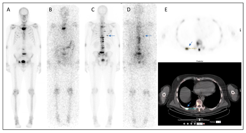
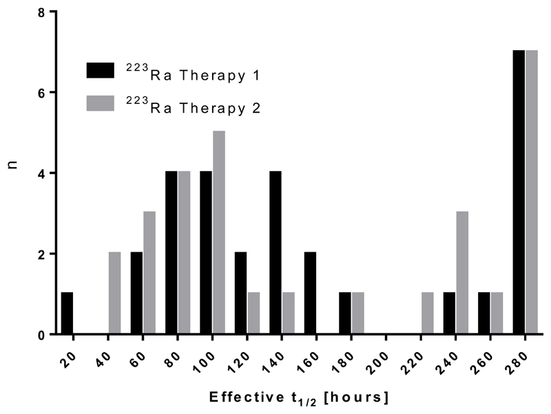
Five patients received two intravenous injections of Ra-dichloride, 6 weeks apart, at 110 kBq/kg whole-body weight. The biodistribution of Ra in metastatic lesions as a function of time after administration as well as associated lesion dosimetry were determined from serial Ra scans. PET/CT imaging using F-fluoride was performed prior to the first treatment (baseline), and at week 6 immediately before the second treatment and at week 12 after baseline.
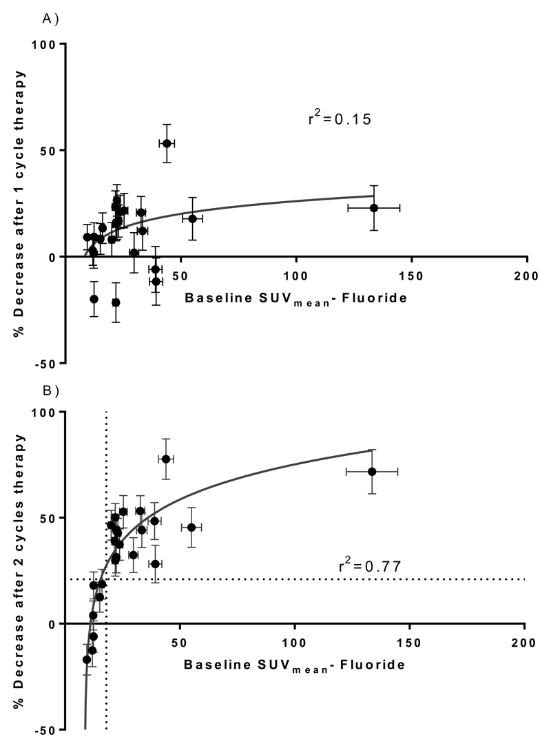
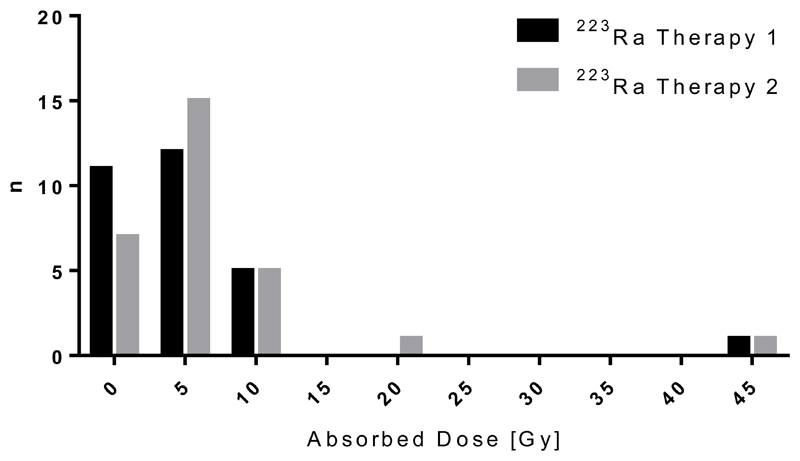
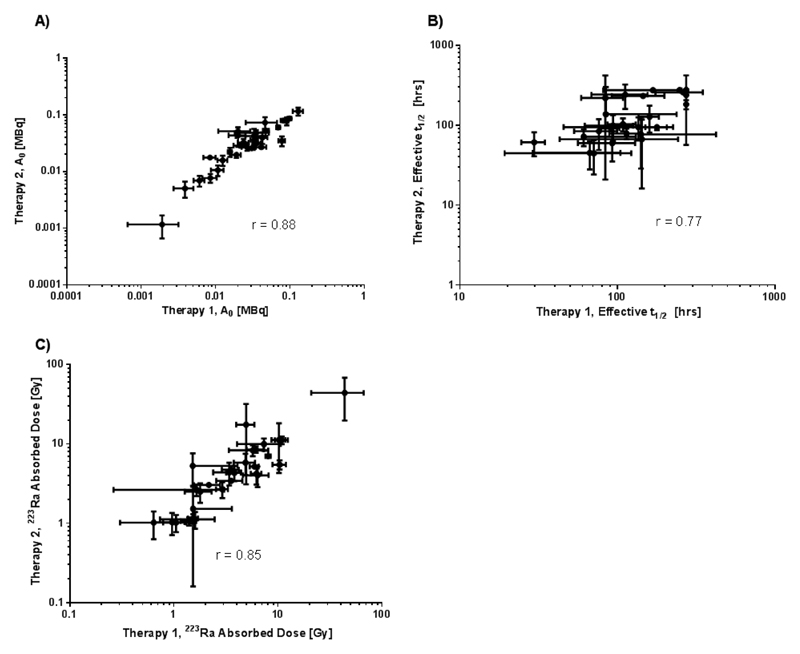
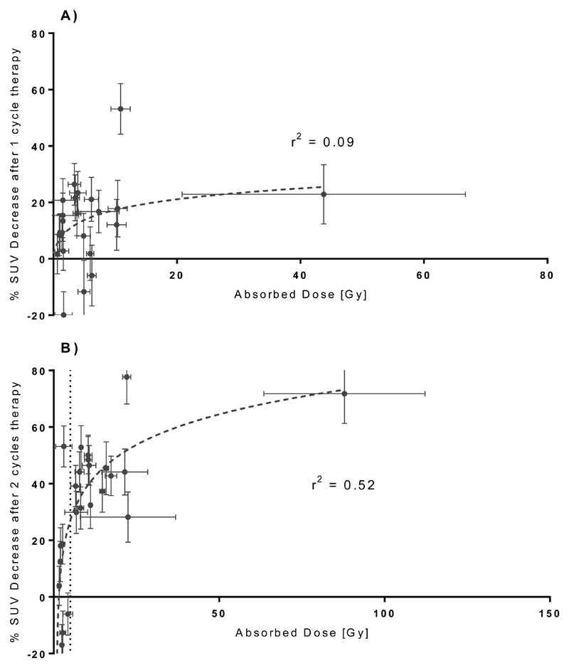
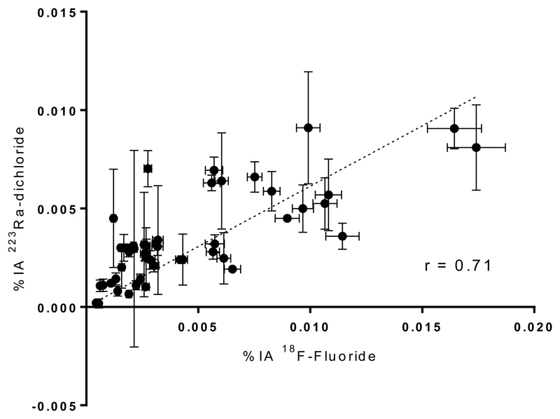
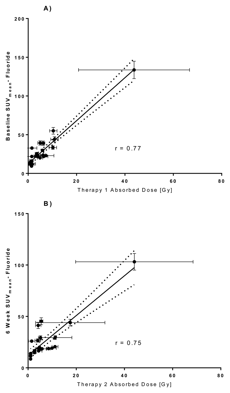
Absorbed doses to metastatic bone lesions ranged from 0.6 Gy to 44.1 Gy. For individual patients, there was an average factor difference of 5.3 (range 2.5-11.0) between the maximum and minimum lesion dose. A relationship between lesion-absorbed doses and serial changes in F-fluoride uptake was demonstrated (r = 0.52). A log-linear relationship was demonstrated (r = 0.77) between baseline measurements of F-fluoride uptake prior to Ra-dichloride therapy and changes in uptake 12 weeks after the first cycle of therapy. Correlations were also observed between both Ra and F-fluoride uptake in lesions (r = 0.75) as well as between Ra absorbed dose and F-fluoride uptake (r = 0.96).
There is both inter-patient and intra-patient heterogeneity of absorbed dose estimates to metastatic lesions. A relationship between Ra lesion absorbed dose and subsequent lesion response was observed. Analysis of this small group of patients suggests that baseline uptake of F-fluoride in bone metastases is significantly correlated with corresponding uptake of Ra, the associated Ra absorbed dose and subsequent lesion response to treatment.
本研究旨在计算基于体重给予镭-二氯化物后骨病变吸收剂量,评估这些剂量与相应 F-氟化物摄取之间的关系,并评估定量 F-氟化物成像预测治疗反应的潜力。
5 名患者在 6 周的间隔内,以 110kBq/kg 全身体重接受两次镭-二氯化物静脉注射。放射性核素扫描确定给药后随时间推移转移病灶中镭的生物分布以及相关病变剂量学。在第一次治疗(基线)前、第二次治疗前的第 6 周和基线后的第 12 周进行 F-氟化物的 PET/CT 成像。
转移性骨病变的吸收剂量范围为 0.6Gy 至 44.1Gy。对于个别患者,最大和最小病变剂量之间的平均差异因子为 5.3(范围为 2.5-11.0)。病变吸收剂量与 F-氟化物摄取的连续变化之间存在关系(r=0.52)。在镭-二氯化物治疗前 F-氟化物摄取的基线测量值与第一个治疗周期后 12 周摄取的变化之间存在对数线性关系(r=0.77)。还观察到病变中镭和 F-氟化物摄取之间的相关性(r=0.75)以及镭吸收剂量与 F-氟化物摄取之间的相关性(r=0.96)。
转移性病变吸收剂量估计存在患者间和患者内异质性。观察到镭病变吸收剂量与随后的病变反应之间存在关系。对这一小组患者的分析表明,骨转移灶中 F-氟化物的基线摄取与相应的镭摄取、相关的镭吸收剂量以及随后的病变对治疗的反应显著相关。