Li Zekun, Benabdallah Nadia, Abou Diane S, Baumann Brian C, Dehdashti Farrokh, Ballard David H, Liu Jonathan, Jammalamadaka Uday, Laforest Richard, Wahl Richard L, Thorek Daniel L J, Jha Abhinav K
Department of Biomedical Engineering, Washington University, St. Louis, MO 63130 USA.
Mallinckrodt Institute of Radiology, Washington University, St. Louis, MO 63110 USA.
IEEE Trans Radiat Plasma Med Sci. 2023 Jan;7(1):62-74. doi: 10.1109/trpms.2022.3175435. Epub 2022 May 23.
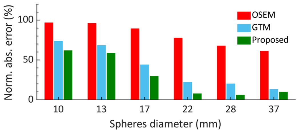
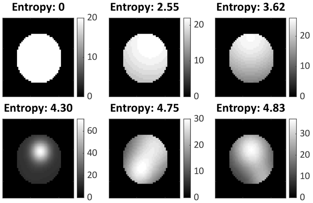
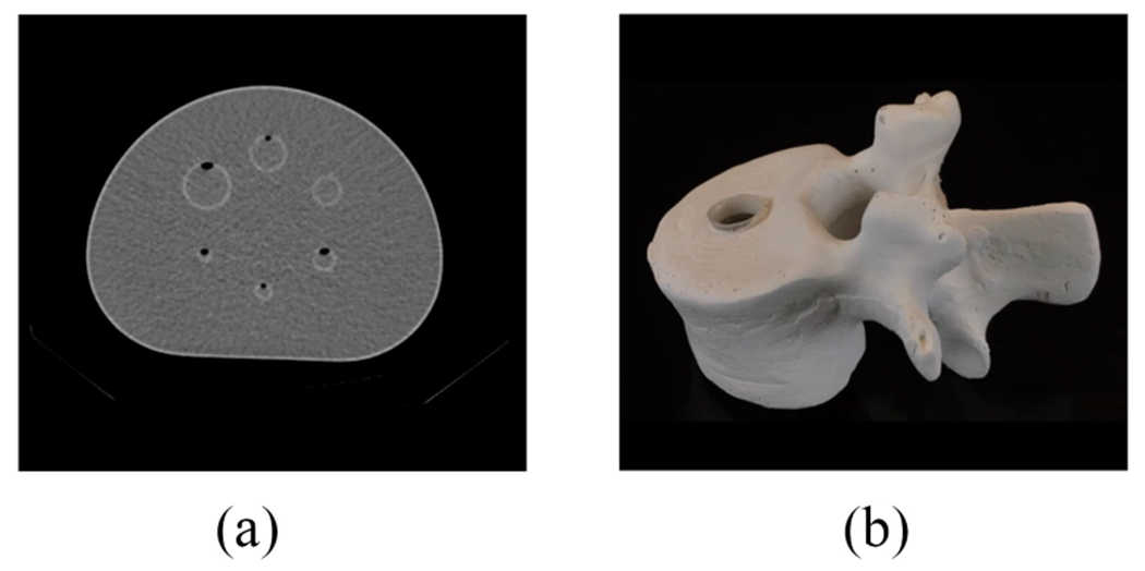
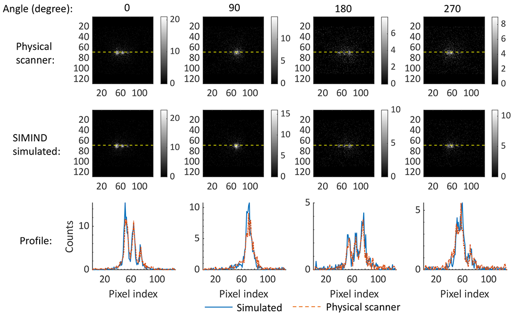
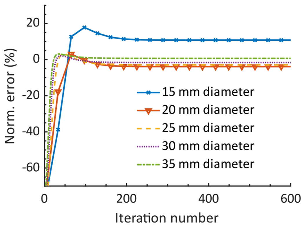
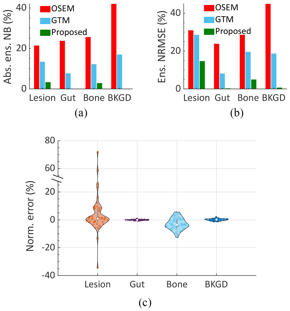
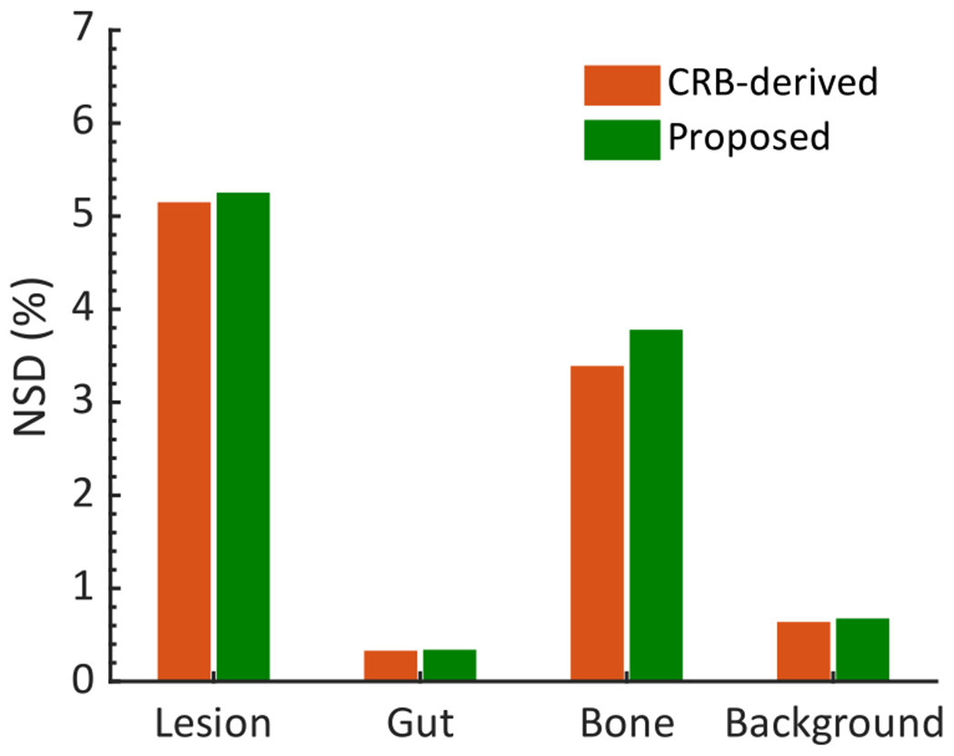
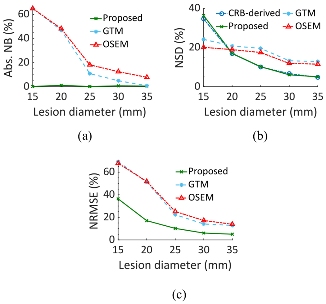
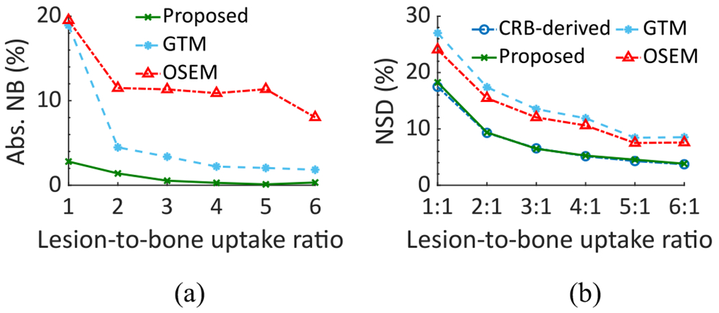
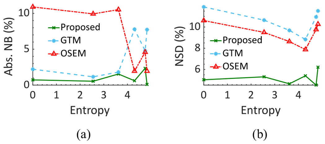
Single-photon emission-computed tomography (SPECT) provides a mechanism to estimate regional isotope uptake in lesions and at-risk organs after administration of -particle-emitting radiopharmaceutical therapies (-RPTs). However, this estimation task is challenging due to the complex emission spectra, the very low number of detected counts (~20 times lower than in conventional SPECT), the impact of stray-radiation-related noise at these low counts, and the multiple image-degrading processes in SPECT. The conventional reconstruction-based quantification methods are observed to be erroneous for -RPT SPECT. To address these challenges, we developed a low-count quantitative SPECT (LC-QSPECT) method that directly estimates the regional activity uptake from the projection data (obviating the reconstruction step), compensates for stray-radiation-related noise, and accounts for the radioisotope and SPECT physics, including the isotope spectra, scatter, attenuation, and collimator-detector response, using a Monte Carlo-based approach. The method was validated in the context of 3-D SPECT with Ra, a commonly used radionuclide for -RPT. Validation was performed using both realistic simulation studies, including a virtual clinical trial, and synthetic and 3-D-printed anthropomorphic physical-phantom studies. Across all studies, the LC-QSPECT method yielded reliable regional-uptake estimates and outperformed the conventional ordered subset expectation-maximization (OSEM)-based reconstruction and geometric transfer matrix (GTM)-based post-reconstruction partial-volume compensation methods. Furthermore, the method yielded reliable uptake across different lesion sizes, contrasts, and different levels of intralesion heterogeneity. Additionally, the variance of the estimated uptake approached the Cramér-Rao bound-defined theoretical limit. In conclusion, the proposed LC-QSPECT method demonstrated the ability to perform reliable quantification for -RPT SPECT.
单光子发射计算机断层扫描(SPECT)提供了一种机制,可在给予发射β粒子的放射性药物治疗(β-RPTs)后,估计病变和高危器官中的区域同位素摄取情况。然而,由于发射光谱复杂、检测到的计数非常少(比传统SPECT低约20倍)、在这些低计数下与杂散辐射相关的噪声影响以及SPECT中的多种图像退化过程,这项估计任务具有挑战性。据观察,传统的基于重建的定量方法对于β-RPT SPECT是错误的。为应对这些挑战,我们开发了一种低计数定量SPECT(LC-QSPECT)方法,该方法直接从投影数据估计区域活性摄取(无需重建步骤),补偿与杂散辐射相关的噪声,并使用基于蒙特卡罗的方法考虑放射性同位素和SPECT物理特性,包括同位素光谱、散射、衰减和准直器-探测器响应。该方法在使用常用的用于β-RPT的放射性核素镭的三维SPECT背景下进行了验证。使用包括虚拟临床试验在内的实际模拟研究以及合成和3D打印的拟人化物理体模研究进行了验证。在所有研究中,LC-QSPECT方法产生了可靠的区域摄取估计,并且优于传统的基于有序子集期望最大化(OSEM)的重建方法和基于几何传递矩阵(GTM)的重建后部分体积补偿方法。此外,该方法在不同病变大小、对比度和不同病变内异质性水平下都能产生可靠的摄取结果。此外,估计摄取的方差接近克拉美-罗下界定义的理论极限。总之,所提出的LC-QSPECT方法证明了对β-RPT SPECT进行可靠定量的能力。