• 文献检索
  • 文档翻译
  • 深度研究
  • 学术资讯
  • Suppr Zotero 插件Zotero 插件
  • 邀请有礼
  • 套餐&价格
  • 历史记录
应用&插件
Suppr Zotero 插件Zotero 插件浏览器插件Mac 客户端Windows 客户端微信小程序
定价
高级版会员购买积分包购买API积分包
服务
文献检索文档翻译深度研究API 文档MCP 服务
关于我们
关于 Suppr公司介绍联系我们用户协议隐私条款
关注我们

Suppr 超能文献

核心技术专利:CN118964589B侵权必究
粤ICP备2023148730 号-1Suppr @ 2026

文献检索

告别复杂PubMed语法,用中文像聊天一样搜索,搜遍4000万医学文献。AI智能推荐,让科研检索更轻松。

立即免费搜索

文件翻译

保留排版,准确专业,支持PDF/Word/PPT等文件格式,支持 12+语言互译。

免费翻译文档

深度研究

AI帮你快速写综述,25分钟生成高质量综述,智能提取关键信息,辅助科研写作。

立即免费体验

慢性猪梗死模型中心血管磁共振成像与高分辨率组织学纤维化定量分析的系统比较

A systematic comparison of cardiovascular magnetic resonance and high resolution histological fibrosis quantification in a chronic porcine infarct model.

作者信息

Gho Johannes M I H, van Es René, van Slochteren Frebus J, Jansen Of Lorkeers Sanne J, Hauer Allard J, van Oorschot Joep W M, Doevendans Pieter A, Leiner Tim, Vink Aryan, Asselbergs Folkert W, Chamuleau Steven A J

机构信息

Department of Cardiology, Division Heart and Lungs, University Medical Center Utrecht, Room E03.511, P.O. Box 85500, 3508 GA, Utrecht, The Netherlands.

Netherlands Heart Institute, Utrecht, The Netherlands.

出版信息

Int J Cardiovasc Imaging. 2017 Nov;33(11):1797-1807. doi: 10.1007/s10554-017-1187-y. Epub 2017 Jun 14.

DOI:10.1007/s10554-017-1187-y
PMID:28616762
原文链接:https://pmc.ncbi.nlm.nih.gov/articles/PMC5682871/
Abstract

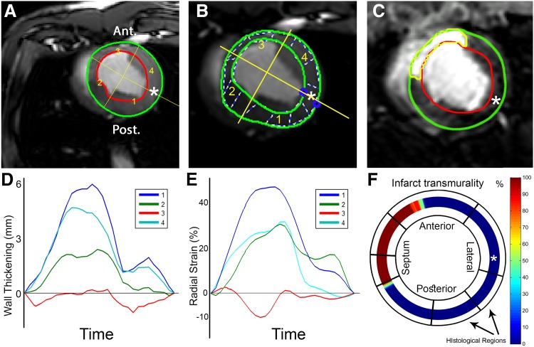
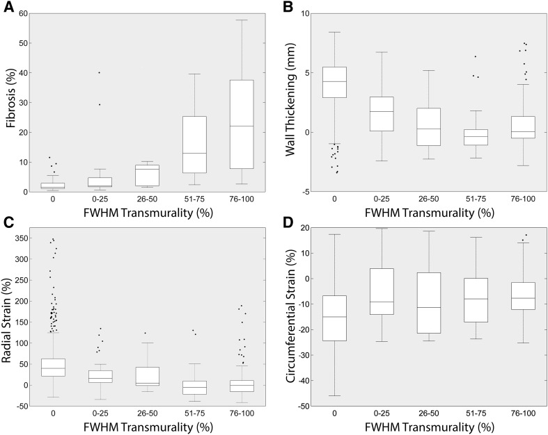
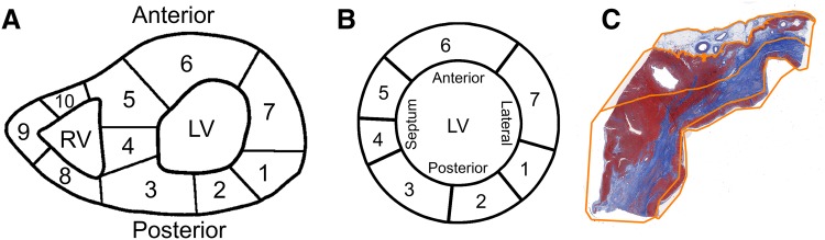
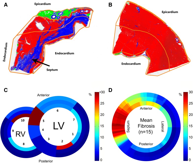
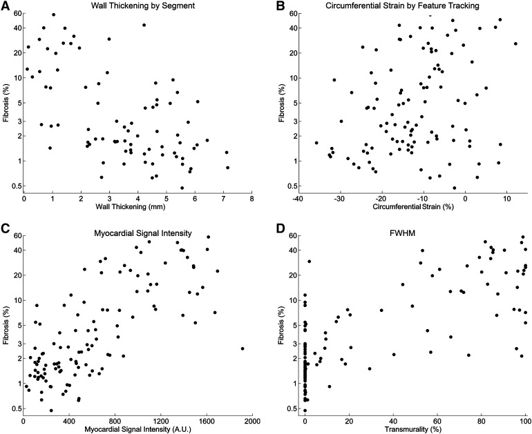
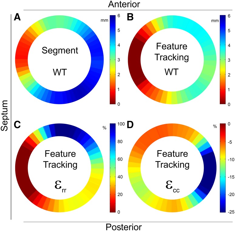
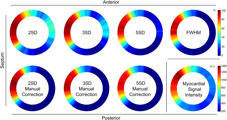
The noninvasive reference standard for myocardial fibrosis detection on cardiovascular magnetic resonance imaging (CMR) is late gadolinium enhancement (LGE). Currently there is no consensus on the preferred method for LGE quantification. Moreover myocardial wall thickening (WT) and strain are measures of regional deformation and function. The aim of this research was to systematically compare in vivo CMR parameters, such as LGE, WT and strain, with histological fibrosis quantification. Eight weeks after 90 min ischemia/reperfusion of the LAD artery, 16 pigs underwent in vivo Cine and LGE CMR. Histological sections from transverse heart slices were digitally analysed for fibrosis quantification. Mean fibrosis percentage of analysed sections was related to the different CMR techniques (using segmentation or feature tracking software) for each slice using a linear mixed model analysis. The full width at half maximum (FWHM) technique for quantification of LGE yielded the highest R of 60%. Cine derived myocardial WT explained 16-36% of the histological myocardial fibrosis. The peak circumferential and radial strain measured by feature tracking could explain 15 and 10% of the variance of myocardial fibrosis, respectively. The used method to systematically compare CMR image data with digital histological images is novel and feasible. Myocardial WT and strain were only modestly related with the amount of fibrosis. The fully automatic FWHM analysis technique is the preferred method to detect myocardial fibrosis.

摘要

心血管磁共振成像(CMR)上检测心肌纤维化的无创参考标准是延迟钆增强(LGE)。目前,关于LGE定量的首选方法尚无共识。此外,心肌壁增厚(WT)和应变是区域变形和功能的指标。本研究的目的是系统地比较体内CMR参数,如LGE、WT和应变,与组织学纤维化定量。在左前降支动脉90分钟缺血/再灌注8周后,16头猪接受了体内电影和LGE CMR检查。对心脏横切片的组织学切片进行数字化分析以进行纤维化定量。使用线性混合模型分析,将分析切片的平均纤维化百分比与每个切片的不同CMR技术(使用分割或特征跟踪软件)相关联。用于LGE定量的半高宽(FWHM)技术产生了最高的60%的R值。电影衍生的心肌WT解释了组织学心肌纤维化的16%-36%。通过特征跟踪测量的峰值圆周应变和径向应变分别可以解释心肌纤维化方差的15%和10%。将CMR图像数据与数字组织学图像进行系统比较的所用方法是新颖且可行的。心肌WT和应变与纤维化程度仅存在适度相关性。全自动FWHM分析技术是检测心肌纤维化的首选方法。

https://cdn.ncbi.nlm.nih.gov/pmc/blobs/b836/5682871/0986ea204803/10554_2017_1187_Fig7_HTML.jpg
https://cdn.ncbi.nlm.nih.gov/pmc/blobs/b836/5682871/0591d519809e/10554_2017_1187_Fig1_HTML.jpg
https://cdn.ncbi.nlm.nih.gov/pmc/blobs/b836/5682871/b5bfd8cde295/10554_2017_1187_Fig2_HTML.jpg
https://cdn.ncbi.nlm.nih.gov/pmc/blobs/b836/5682871/6ea9754c0118/10554_2017_1187_Fig3_HTML.jpg
https://cdn.ncbi.nlm.nih.gov/pmc/blobs/b836/5682871/678dadb59396/10554_2017_1187_Fig4_HTML.jpg
https://cdn.ncbi.nlm.nih.gov/pmc/blobs/b836/5682871/1d2b95db359c/10554_2017_1187_Fig5_HTML.jpg
https://cdn.ncbi.nlm.nih.gov/pmc/blobs/b836/5682871/bdbfd62721e3/10554_2017_1187_Fig6_HTML.jpg
https://cdn.ncbi.nlm.nih.gov/pmc/blobs/b836/5682871/0986ea204803/10554_2017_1187_Fig7_HTML.jpg

相似文献

1
A systematic comparison of cardiovascular magnetic resonance and high resolution histological fibrosis quantification in a chronic porcine infarct model.慢性猪梗死模型中心血管磁共振成像与高分辨率组织学纤维化定量分析的系统比较
Int J Cardiovasc Imaging. 2017 Nov;33(11):1797-1807. doi: 10.1007/s10554-017-1187-y. Epub 2017 Jun 14.
2
Validation of contrast enhanced cine steady-state free precession and T2-weighted CMR for assessment of ischemic myocardial area-at-risk in the presence of reperfusion injury.在存在再灌注损伤的情况下,对比增强电影稳态自由进动和T2加权心脏磁共振成像用于评估缺血心肌危险区域的验证。
Int J Cardiovasc Imaging. 2019 Jun;35(6):1039-1045. doi: 10.1007/s10554-019-01569-x. Epub 2019 Mar 9.
3
Comparison of fast multi-slice and standard segmented techniques for detection of late gadolinium enhancement in ischemic and non-ischemic cardiomyopathy - a prospective clinical cardiovascular magnetic resonance trial.比较快速多层面和标准分段技术在缺血性和非缺血性心肌病中的延迟钆增强检测 - 一项前瞻性临床心血管磁共振试验。
J Cardiovasc Magn Reson. 2018 Feb 19;20(1):13. doi: 10.1186/s12968-018-0434-2.
4
Assessment of myocardial injury after reperfused infarction by T1ρ cardiovascular magnetic resonance.通过T1ρ心血管磁共振评估再灌注性心肌梗死后的心肌损伤
J Cardiovasc Magn Reson. 2017 Feb 15;19(1):17. doi: 10.1186/s12968-017-0332-z.
5
Three-dimensional regional strain analysis in porcine myocardial infarction: a 3T magnetic resonance tagging study.猪心肌梗死的三维区域性应变分析:3T 磁共振标记研究。
J Cardiovasc Magn Reson. 2012 Dec 13;14(1):85. doi: 10.1186/1532-429X-14-85.
6
In vivo contrast free chronic myocardial infarction characterization using diffusion-weighted cardiovascular magnetic resonance.使用扩散加权心血管磁共振对体内无对比剂的慢性心肌梗死进行特征描述。
J Cardiovasc Magn Reson. 2014 Sep 17;16(1):68. doi: 10.1186/s12968-014-0068-y.
7
Patterns of CMR measured longitudinal strain and its association with late gadolinium enhancement in patients with cardiac amyloidosis and its mimics.心脏淀粉样变性及其模仿疾病患者中CMR测量的纵向应变模式及其与延迟钆增强的关联。
J Cardiovasc Magn Reson. 2017 Aug 7;19(1):61. doi: 10.1186/s12968-017-0376-0.
8
Native T1 mapping to detect extent of acute and chronic myocardial infarction: comparison with late gadolinium enhancement technique.利用天然T1映射检测急性和慢性心肌梗死范围:与延迟钆增强技术的比较
Int J Cardiovasc Imaging. 2019 Mar;35(3):517-527. doi: 10.1007/s10554-018-1467-1. Epub 2018 Oct 24.
9
Feasibility of detecting myocardial infarction in the sheep fetus using late gadolinium enhancement CMR imaging.应用钆延迟增强磁共振成像检测绵羊胎儿心肌梗死的可行性。
J Cardiovasc Magn Reson. 2017 Sep 13;19(1):69. doi: 10.1186/s12968-017-0383-1.
10
Three-dimensional speckle tracking longitudinal strain is related to myocardial fibrosis determined by late-gadolinium enhancement.三维斑点追踪纵向应变与延迟钆增强测定的心肌纤维化相关。
Int J Cardiovasc Imaging. 2017 Sep;33(9):1351-1360. doi: 10.1007/s10554-017-1115-1. Epub 2017 Mar 15.

引用本文的文献

1
Quantitative Analyses of the Left Ventricle Volume and Cardiac Function in Normal and Infarcted Yucatan Minipigs.正常和梗死的尤卡坦小型猪左心室容积和心脏功能的定量分析
J Imaging. 2021 Jul 1;7(7):107. doi: 10.3390/jimaging7070107.
2
Feature tracking strain analysis detects the onset of regional diastolic dysfunction in territories with acute myocardial injury induced by transthoracic electrical interventions.特征跟踪应变分析可检测经胸电干预引起的急性心肌损伤区域的局部舒张功能障碍的发生。
Sci Rep. 2022 Nov 14;12(1):19532. doi: 10.1038/s41598-022-24199-1.
3
3D Myocardial Scar Prediction Model Derived from Multimodality Analysis of Electromechanical Mapping and Magnetic Resonance Imaging.

本文引用的文献

1
Comparison of cardiovascular magnetic resonance feature tracking and tagging for the assessment of left ventricular systolic strain in acute myocardial infarction.心血管磁共振特征追踪与标记在急性心肌梗死左心室收缩期应变评估中的比较
Eur J Radiol. 2015 May;84(5):840-8. doi: 10.1016/j.ejrad.2015.02.002. Epub 2015 Feb 16.
2
Image Feature Determinants of Global and Segmental Circumferential Ventricular Strain From Cine CMR.来自心脏磁共振电影成像的整体和节段性圆周心室应变的图像特征决定因素
JACC Cardiovasc Imaging. 2015 Dec;8(12):1465-1466. doi: 10.1016/j.jcmg.2014.10.005. Epub 2014 Nov 1.
3
Histological validation of cardiac magnetic resonance analysis of regional and diffuse interstitial myocardial fibrosis.
基于机电图与磁共振成像多模态分析的三维心肌瘢痕预测模型。
J Cardiovasc Transl Res. 2019 Dec;12(6):517-527. doi: 10.1007/s12265-019-09899-w. Epub 2019 Jul 23.
4
Multimodality imaging for real-time image-guided left ventricular lead placement during cardiac resynchronization therapy implantations.心脏再同步治疗植入术中用于实时影像引导左心室导线置入的多模态成像
Int J Cardiovasc Imaging. 2019 Jul;35(7):1327-1337. doi: 10.1007/s10554-019-01574-0. Epub 2019 Mar 7.
5
Validation of a novel stand-alone software tool for image guided cardiac catheter therapy.一种用于图像引导心脏导管治疗的新型独立软件工具的验证
Int J Cardiovasc Imaging. 2019 Feb;35(2):225-235. doi: 10.1007/s10554-019-01541-9. Epub 2019 Jan 28.
6
Cardiovascular imaging 2017 in the International Journal of Cardiovascular Imaging.《国际心血管影像杂志》2017年心血管成像相关内容
Int J Cardiovasc Imaging. 2018 Jun;34(6):833-848. doi: 10.1007/s10554-018-1349-6.
心肌磁共振分析区域性和弥漫性间质纤维化的组织学验证。
Eur Heart J Cardiovasc Imaging. 2015 Jan;16(1):14-22. doi: 10.1093/ehjci/jeu182. Epub 2014 Oct 28.
4
Myocardial infarction and functional outcome assessment in pigs.猪的心肌梗死与功能结局评估
J Vis Exp. 2014 Apr 25(86):51269. doi: 10.3791/51269.
5
High resolution systematic digital histological quantification of cardiac fibrosis and adipose tissue in phospholamban p.Arg14del mutation associated cardiomyopathy.在受磷蛋白p.Arg14del突变影响的心肌病中对心脏纤维化和脂肪组织进行高分辨率系统性数字组织学定量分析。
PLoS One. 2014 Apr 14;9(4):e94820. doi: 10.1371/journal.pone.0094820. eCollection 2014.
6
Feature tracking compared with tissue tagging measurements of segmental strain by cardiovascular magnetic resonance.心血管磁共振特征追踪与组织标记法测量节段应变的比较。
J Cardiovasc Magn Reson. 2014 Jan 22;16(1):10. doi: 10.1186/1532-429X-16-10.
7
Validation of contrast-enhanced magnetic resonance imaging to monitor regenerative efficacy after cell therapy in a porcine model of convalescent myocardial infarction.在猪心肌梗死后恢复期细胞治疗模型中,使用对比增强磁共振成像监测再生疗效的验证。
Circulation. 2013 Dec 24;128(25):2764-75. doi: 10.1161/CIRCULATIONAHA.113.002863. Epub 2013 Sep 23.
8
Quantification of fibrosis in infarcted swine hearts by ex vivo late gadolinium-enhancement and diffusion-weighted MRI methods.应用离体晚期钆增强和弥散加权 MRI 方法对梗死猪心纤维化进行定量评估。
Phys Med Biol. 2013 Aug 7;58(15):5009-28. doi: 10.1088/0031-9155/58/15/5009. Epub 2013 Jul 8.
9
Global and regional left ventricular myocardial deformation measures by magnetic resonance feature tracking in healthy volunteers: comparison with tagging and relevance of gender.健康志愿者心脏磁共振特征追踪技术检测的左心室心肌整体和局部形变:与标记法的比较及与性别相关。
J Cardiovasc Magn Reson. 2013 Jan 18;15(1):8. doi: 10.1186/1532-429X-15-8.
10
The intra-observer reproducibility of cardiovascular magnetic resonance myocardial feature tracking strain assessment is independent of field strength.心血管磁共振心肌特征追踪应变评估的观察者内可重复性与磁场强度无关。
Eur J Radiol. 2013 Feb;82(2):296-301. doi: 10.1016/j.ejrad.2012.11.012. Epub 2012 Dec 12.