Ekino Taisuke, Yoshiga Toyoshi, Takeuchi-Kaneko Yuko, Kanzaki Natsumi
Laboratory of Nematology, Department of Applied Biological Sciences, Faculty of Agriculture, Saga University, Saga, Japan.
The United Graduate School of Agricultural Sciences, Kagoshima University, Kagoshima, Japan.
PLoS One. 2017 Jun 16;12(6):e0179465. doi: 10.1371/journal.pone.0179465. eCollection 2017.
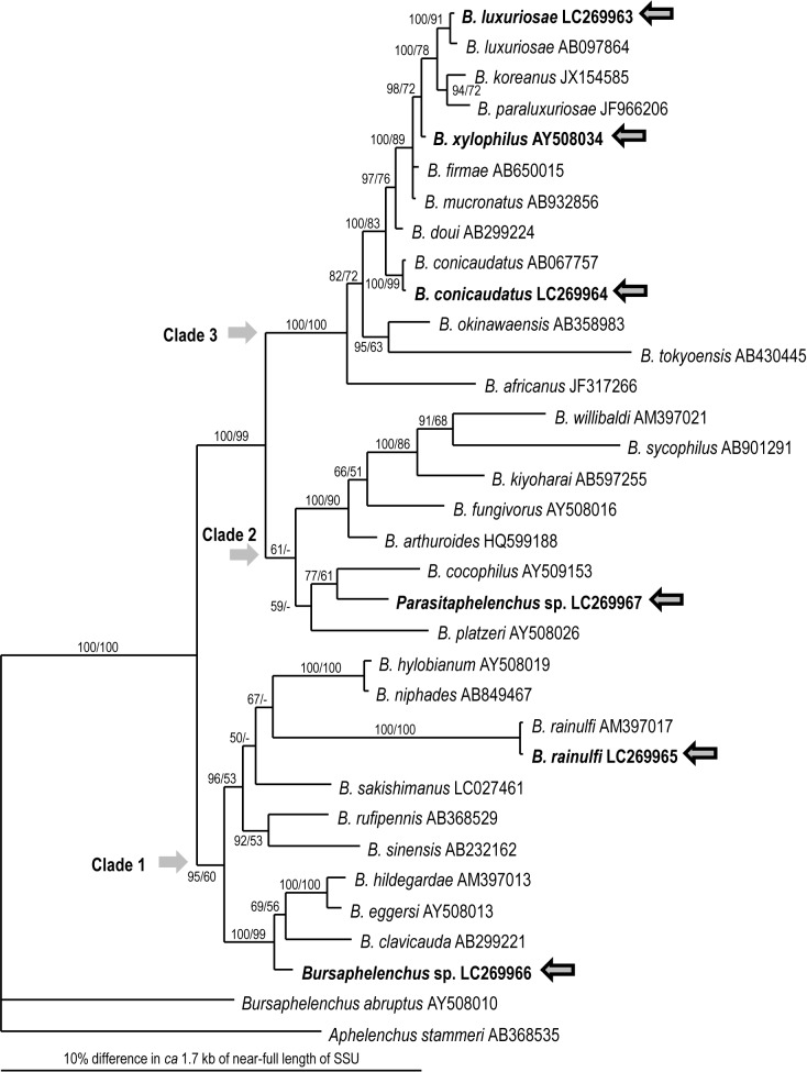
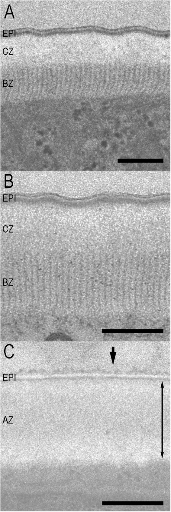
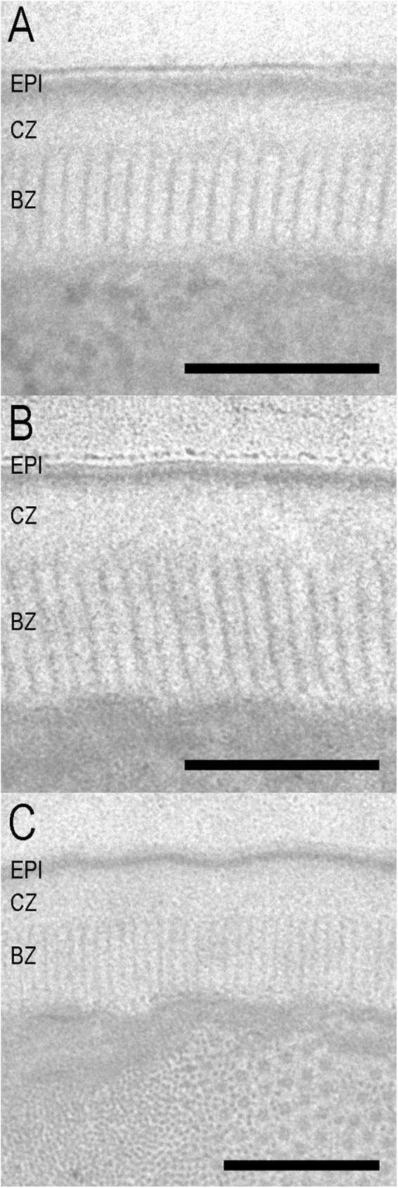
Using transmission electron microscopy, we examined the body cuticle ultrastructures of phoretic and parasitic stages of the parasitaphelenchid nematodes Bursaphelenchus xylophilus, B. conicaudatus, B. luxuriosae, B. rainulfi; an unidentified Bursaphelenchus species, and an unidentified Parasitaphelenchus species. Nematode body cuticles usually consist of three zones, a cortical zone, a median zone, and a basal zone. The phoretic stages of Bursaphelenchus spp., isolated from the tracheal systems of longhorn beetles or the elytra of bark beetles, have a thick and radially striated basal zone. In contrast, the parasitic stage of Parasitaphelenchus sp., isolated from bark beetle hemocoel, has no radial striations in the basal zone. This difference probably reflects the peculiar ecological characteristics of the phoretic stage. A well-developed basal radially striated zone, composed of very closely linked proteins, is the zone closest to the body wall muscle. Therefore, the striation is necessary for the phoretic species to be able to seek, enter, and depart from host/carrier insects, but is not essential for internal parasites in parasitaphelenchid nematodes. Phylogenetic relationships inferred from near-full-length small subunit ribosomal RNA sequences suggest that the cuticle structures of parasitic species have apomorphic characters, e.g., lack of striation in the basal zone, concurrent with the evolution of insect parasitism from a phoretic life history.
我们使用透射电子显微镜检查了松材线虫(Bursaphelenchus xylophilus)、拟松材线虫(B. conicaudatus)、华丽松材线虫(B. luxuriosae)、雷氏松材线虫(B. rainulfi)、一种未鉴定的松材线虫物种以及一种未鉴定的拟寄生线虫物种的携播阶段和寄生阶段的体表角质层超微结构。线虫的体表角质层通常由三个区域组成,即皮层区、中区和基部区。从天牛气管系统或小蠹虫鞘翅分离得到的松材线虫属的携播阶段,其基部区厚且有径向条纹。相比之下,从小蠹虫血腔分离得到的拟寄生线虫属的寄生阶段,其基部区没有径向条纹。这种差异可能反映了携播阶段独特的生态特征。一个由紧密相连的蛋白质组成的发育良好的基部径向条纹区,是最靠近体壁肌肉的区域。因此,这种条纹对于携播物种寻找、进入和离开宿主/载体昆虫是必要的,但对于拟寄生线虫中的内寄生线虫来说并非必不可少。从近乎全长的小亚基核糖体RNA序列推断的系统发育关系表明,寄生物种的角质层结构具有衍生特征,例如基部区缺乏条纹,这与从携播生活史向昆虫寄生的进化同时发生。