Grönman Maria, Tarkia Miikka, Kiviniemi Tuomas, Halonen Paavo, Kuivanen Antti, Savunen Timo, Tolvanen Tuula, Teuho Jarmo, Käkelä Meeri, Metsälä Olli, Pietilä Mikko, Saukko Pekka, Ylä-Herttuala Seppo, Knuuti Juhani, Roivainen Anne, Saraste Antti
Turku PET Centre, University of Turku, 20521, Turku, Finland.
Heart Center, Turku University Hospital, Turku, Finland.
J Transl Med. 2017 Jun 19;15(1):144. doi: 10.1186/s12967-017-1245-1.
Radiolabeled RGD peptides detect αβ integrin expression associated with angiogenesis and extracellular matrix remodeling after myocardial infarction. We studied whether cardiac positron emission tomography (PET) with [Ga]NODAGA-RGD detects increased αβ integrin expression after induction of flow-limiting coronary stenosis in pigs, and whether αβ integrin is expressed in viable ischemic or injured myocardium.
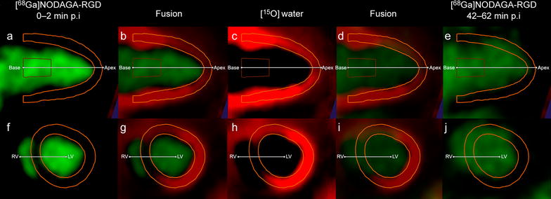
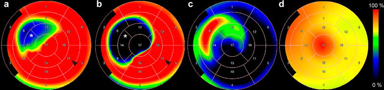
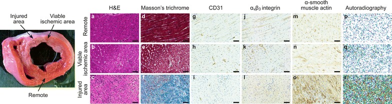
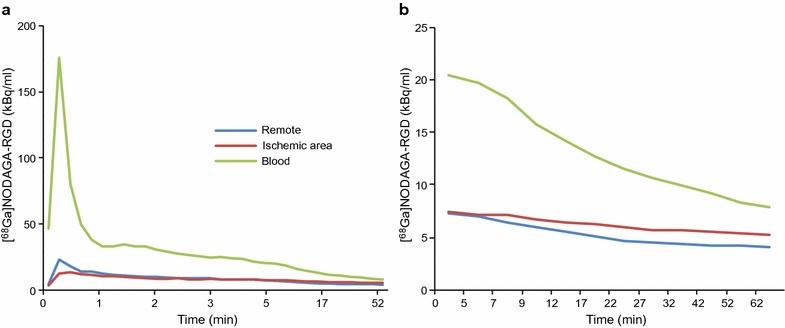
We studied 8 Finnish landrace pigs 13 ± 4 days after percutaneous implantation of a bottleneck stent in the proximal left anterior descending coronary artery. Antithrombotic therapy was used to prevent stent occlusion. Myocardial uptake of [Ga]NODAGA-RGD (290 ± 31 MBq) was evaluated by a 62 min dynamic PET scan. The ischemic area was defined as the regional perfusion abnormality during adenosine-induced stress by [O]water PET. Guided by triphenyltetrazolium chloride staining, tissue samples from viable and injured myocardial areas were obtained for autoradiography and histology.
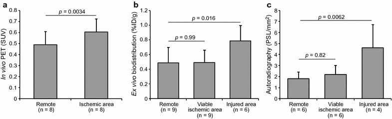
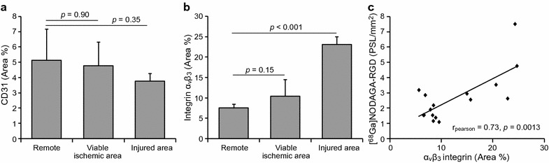
Stent implantation resulted in a partly reversible myocardial perfusion abnormality. Compared with remote myocardium, [Ga]NODAGA-RGD PET showed increased tracer uptake in the ischemic area (ischemic-to-remote ratio 1.3 ± 0.20, p = 0.0034). Tissue samples from the injured areas, but not from the viable ischemic areas, showed higher [Ga]NODAGA-RGD uptake than the remote non-ischemic myocardium. Uptake of [Ga]NODAGA-RGD correlated with immunohistochemical detection of αβ integrin that was expressed in the injured myocardial areas.
Cardiac [Ga]NODAGA-RGD PET demonstrates increased myocardial αβ integrin expression after induction of flow-limiting coronary stenosis in pigs. Localization of [Ga]NODAGA-RGD uptake indicates that it reflects αβ integrin expression associated with repair of recent myocardial injury.
放射性标记的RGD肽可检测与心肌梗死后血管生成和细胞外基质重塑相关的αβ整合素表达。我们研究了用[Ga]NODAGA-RGD进行心脏正电子发射断层扫描(PET)是否能检测猪在诱导限流性冠状动脉狭窄后αβ整合素表达的增加,以及αβ整合素是否在存活的缺血或损伤心肌中表达。
我们对8只芬兰长白猪进行研究,在经皮将瓶颈支架植入左冠状动脉前降支近端13±4天后进行。采用抗血栓治疗以防止支架闭塞。通过62分钟的动态PET扫描评估[Ga]NODAGA-RGD(290±31MBq)的心肌摄取情况。缺血区域通过[O]水PET在腺苷诱导的应激期间的区域灌注异常来定义。在氯化三苯基四氮唑染色的引导下,从存活和损伤心肌区域获取组织样本用于放射自显影和组织学检查。
支架植入导致部分可逆的心肌灌注异常。与远隔心肌相比,[Ga]NODAGA-RGD PET显示缺血区域的示踪剂摄取增加(缺血与远隔比值为1.3±0.20,p = 0.0034)。来自损伤区域而非存活缺血区域的组织样本显示,[Ga]NODAGA-RGD摄取高于远隔非缺血心肌。[Ga]NODAGA-RGD的摄取与在损伤心肌区域表达的αβ整合素的免疫组织化学检测相关。
心脏[Ga]NODAGA-RGD PET显示猪在诱导限流性冠状动脉狭窄后心肌αβ整合素表达增加。[Ga]NODAGA-RGD摄取的定位表明它反映了与近期心肌损伤修复相关的αβ整合素表达。